More Than Sick of Salt

All Posts in Category: Gastroparesis

GASTROPARESIS LIKE SYNDROME AND DUMPING SYNDROME

DUMPING SYNDROME AND GASTROPARESIS-LIKE SYNDROME

Click here to download this post

Gastroparesis is a chronic disease accompanied by bloating, early fullness after a meal, nausea, vomiting, and abdominal pain.  A diagnosis of Gastroparesis requires objective data demonstrating delayed gastric emptying in the absence of intestinal obstruction.  There are, however, disorders known as Gastroparesis-Like Syndrome (GLS).  Patients with GLS have the same or similar symptoms to those who have Gastroparesis, but on gastric emptying studies it shows normal or rapid emptying.  Interestingly, both patient types, those with Gastroparesis (slowed or delayed emptying) and those with normal or rapid emptying, have been shown to benefit from Gastro-Electrical Stimulation (GES) placement.  GLS may be actually be a spectrum of Gastroparesis.  Patients who present with unexplained nausea and vomiting for at least 12 weeks without evidence of obstruction should be evaluated for both Gastroparesis and GLS.  Chronic unexplained nausea patients may have similar abnormal scores on the Gastroparesis Cardinal Symptom Index (GCSI); therefore, an objective test of gastric emptying needs to be done, such as a Scintigraphic Gastric Emptying study.  For patients who have chronic unexplained nausea and vomiting on biopsy have loss of neuronal Nitric Oxide Synthase (NOS) and loss of Interstitial Cells of Cajal.  These two findings are also seen in chronic Gastroparesis.

As a quick summary, when comparing patients with Gastroparesis with delayed gastric emptying and GLS with normal or rapid emptying, they both have similar GSCI scores.  The difference is in the gastric emptying study.  Gastroparesis patients appear to have slow emptying and a loss of the Interstitial Cells of Cajal.  However, the Cells of Cajal also decrease in GLS patients.

Inflammation has been found in patients with these disorders, usually with elevated C-Reactive Protein (CRP) levels.  CRP is produced in the liver and responds sensitively to inflammation, making it a very good marker of inflammation.  Inflammation, as indicated by elevated CRP, is typically very prevalent in Diabetic Gastroparesis.  One study showed that patients treated with immunomodulating agents, such as IVIG or Mycophenolate Mofetil or a combination of steroids and Mycophenolate Mofetil improved in 8-12 weeks supporting an anti-inflammatory and possibility and autoimmune mechanism.  Studies confirm the hypothesis that patients with Gastroparesis and GLS are part of a spectrum and inflammation is an underlying factor.

Prospective studies are needed to further assess changes in the Autonomic Nervous System (ANS), Central Nervous System and Enteric pathways that play a role in Pyloric, Antrum and Gastric emptying.  Rapid emptying may present in the form of a dumping type syndrome.  While this is a common complication of esophageal, gastric or bariatric surgery and may include both early and late dumping components, it also can be seen in idiopathic states and post viral.  Liquid meals may be better to detect acceleration of early gastric emptying than in solid meals.  Solid meals generally have a low sensitivity and specificity for detecting accelerated gastric emptying.  Therefore, if someone is suspected of having dumping syndrome and has a normal gastric emptying study with a solid meal, a liquid meal should be considered.

Most people with dumping syndrome develop signs and symptoms, such as abdominal cramps and diarrhea 10-30 minutes after eating while other people can just have symptoms three hours later, which includes symptoms of hypoglycemia.  Generally, the early symptoms result when a patient feels bloated or too full after eating.  That is early satiety.  Also, nausea, vomiting, abdominal cramps, diarrhea, flushing, dizziness, lightheadedness, and rapid heart rate can be experienced.  Late dumping syndrome starts one to three hours after a meal, especially one that is high in sugar.  The dumping is usually a hypoglycemic or low sugar abnormality.  It is usually due to the release of a large amount of insulin to absorb the sugars entering the small intestine.  These can produce vasomotor (vascular) symptoms, including sweating and flushing, lightheadedness, weakness and rapid heart rate (palpitation), plus an intense desire to lie down.  Physical exam of these patients show significant orthostatic changes, not just increase in heart rate but also a drop in blood pressure can occur upon standing or sitting-up.  An abnormal change in blood pressure (including a decrease of any sort) upon assuming an upright posture (sitting up or standing) is known as Orthostatic Intolerance, and in the more extreme cases Orthostatic Hypotension.  Vasomotor symptoms predominate.  The late dumping symptom of orthostatic dysfunction is a consequence of active hypoglycemia from exaggerated release of insulin.

Some patients do not experience a drop in blood pressure when assuming an upright posture, rather they experience a rapid or irregular heart rate (tachycardia or palpitations).  Occasionally Propranolol, or a low-dose Verapamil, is useful in treating these rapid or irregular heartbeats which can occur in dumping syndrome.  These abnormal heart rate patterns are characteristic of the orthostatic dysfunction of Postural Orthostatic Tachycardia Syndrome (POTS).  POTS tends to occur more in younger females.  This is due to the fact that women are born with hearts that are physically smaller in size (especially thinner muscle walls) than men.  This may cause women to experience cardiac deconditioning more than men, and in these cases, since the heart is smaller, it cannot leverage pressure; therefore, it leverages rate in its attempt to deliver more blood to the brain.  This can occur in the early or late stage gastric dumping.

Most people that develop dumping syndrome have early dumping and only about a quarter have late dumping.  The early dumping patients generally have both abdominal symptoms and vasomotor symptoms.  The abdominal symptoms, as mentioned, include early satiety, abdominal pains, nausea, cramps, diarrhea and vomiting, whereas the vasomotor symptoms include the sweating, flushing, tachycardia, palpitations, low blood pressure, headaches and at times even passing out, or syncope.  These symptoms are related to the bowel becoming distended and hormones being secreted by the GI tract and activation of the ANS, specifically the Parasympathetic Nervous System.  Therefore, with early dumping, one has both vasomotor and gastrointestinal symptoms.  One to three hours later, the second phase, one has reactive low blood sugar or reactive hypoglycemia symptoms.  These are predominately what we call vasomotor in origin.  If a patient develops dumping syndrome, they often avoid food and eating because symptoms are so uncomfortable.

Usually, an oral glucose challenge of 50 grams of glucose is given.  An increased heart rate by 10 beats per minute in the first hour is considered a positive test.  Also, if the hematocrit increases 3% in the first 30 minutes that suggests dumping syndrome.  Late dumping syndrome is indicated if one develops low blood sugar 2 to 3 hours later.  Radionuclide Scintography, also known as Gastric Scintography, demonstrates rapid gastric emptying with standardized tests.  The main mechanism for dumping syndrome is the rapid introduction of partially digested food into the small intestine.

Dumping syndrome is often seen after gastroesophageal surgeries such as Fundoplication (a surgical procedure to treat gastric reflux), or bariatric surgeries.  It had at one time been seen for surgical treatment of peptic ulcer disease.  This is rarely the case since medical therapy is very effective.  Gastric bypass is the most common cause of dumping syndrome that we see today in adults.  Up to 75% of patients have dumping syndrome after gastric surgery, but many of them learn how to handle this with proper dietary intake.  Dietary modifications are important.  Complex carbohydrates, small meals of six per day, reducing the actual carbohydrate quantity, and fluid intake are important to modify.  Fluids should be taken one hour after meals or after ingestion of solids, since liquids will quicken the transit time through the stomach.  Dairy products should be avoided.  Fats and proteins are preferred over carbohydrates.  Increasing dietary fiber helps to treat the reactive hypoglycemia that is seen in a delayed response and it also slows the gastric emptying.  If one feels lightheaded and they have low blood pressure they should lie down after eating.

If a patient does not respond to dietary moderation, low doses of Loperamide may be beneficial for the diarrhea.  Candy is useful to relieve the hypoglycemia which may occur later.  Oftentimes, abdominal distention and bloating can be controlled with probiotics.   A medicine used in treating diabetes, Acarbose, has been useful for the late stage of dumping syndrome.  It lowers the blood sugar elevation after eating and helps to control reactive hypoglycemia.  Anticholinergic medicines also may be very useful in slowing rapid GI transit due to Parasympathetic Excess.  Antispasm medicines, such as Dicyclomine (Benadryl) or Propantheline, may helpful.  There are other more advanced medicines which Gastroenterologists can use such as Diazoxide to control the reactive hypoglycemia in the late dumping stage.  Another advanced medication is Somatostatin, but these advanced medications are used in patients with intractable symptoms.  GES is a recently developed advanced treatment.  Interestingly, GES normalizes gastric transit time by actually slowing it, and patients respond.  This should be considered, just as in Gastroparesis, in patients that are unresponsive to dietary changes and drug therapy.  GES helps improve rapid gastric emptying and causes increased gastric retention of food and reduces nausea and vomiting.

Many times the Dumping Syndrome is not diagnosed.  It can be seen in Diabetes, but there is also an idiopathic form and surgery need not necessarily be present.  Rumination Syndrome and Cyclical Vomiting Syndrome need to be excluded.  Rumination Syndrome is an effortless regurgitation of gastric contents into the mouth, caused by contraction of the abdominal wall, and subsequent re-swallowing of food.  A large number of patients who have not had gastric surgery have underlying Anxiety, Depression or Diabetes Mellitus, and a few have a diagnosis of Cyclic Vomiting Syndrome or reported Cannabis use.  Cannabis may cause rapid gastric emptying and a condition known as Cannabinoid Hyperemesis.  Cannabis may also cause unexplained upper gastrointestinal symptoms, as seen in some hospitalized patients.

Patients without prior history of gastric surgery that have dumping syndrome are classified as idiopathic.  In the past, we diagnosed these people as having non-ulcer dyspepsia.  However, the patients who have dumping syndrome usually have more severe abdominal cramping as well as systemic symptoms of sweating, weakness, palpitations, flushing and dizziness which is more pronounced in people with just non-ulcer dyspepsia.  About a third of these patients with Idiopathic Dumping Syndrome have had a prior gastroenteritis probably due to a viral mechanism.  It is believed that injury to the duodenal receptors, mainly the osmotic and fat receptors, which control gastric emptying, may be damaged.  Another is that the Vagus Nerve (a major part of the Parasympathetic nervous system) is only partially damaged.  It is speculated that rapid gastric emptying is due to early Vagal damage where just the distal end of the Vagus Nerve is damaged.  This is a part of a spectrum.  When the entire Vagus Nerve is damaged, one could get slow gastric transit or Gastroparesis.  Gastroparesis is a possible state that evolves into a more complete Vagal loss (as in Diabetes).  The Vagus Nerve is the longest nerve in the body and is very susceptible to damage both at surgery and viral infections as well as inflammation or oxidative stress (stress at the cellular level).

Among the identifiable causative factors for dumping syndrome in nonsurgical patients, Diabetes is the most common.  Therefore, Diabetes can cause both (1) a rapid emptying or a dumping-type presentation, as well as, (2) a delayed emptying or a Gastroparesis-type presentation.  Therefore, with significant abdominal symptoms with nausea and vomiting, especially if they have vasomotor symptoms, dumping syndrome should be suspected, although the symptoms may be identical to Gastroparesis and a gastric emptying study will differentiate the two.  This is important because treatment and pharmacology will differ.  In all refractory patients, GES by different mechanisms may improve the gastric motility.  Rarely are surgical revisions necessary in patients with dumping syndrome except if they have had prior bariatric surgery, in which case many times revisions might be indicated.

Rapid gastric emptying sometimes occurs in people who have not had stomach surgery.  For example, those who have recent onset of Diabetes, especially type 2 Diabetes.  This is in contrast to Gastroparesis which usually is a late finding in Diabetes and more often seen in Diabetes type 2 than type 1.  Non-surgical, rapid emptying may also occur in patients with (1) Pancreatic exocrine insufficiency, which can cause problems with digestion, or (2) Duodenal ulcers, or (3) Zollinger-Ellison syndrome, which is a rare condition in which one or more tumors form in your pancreas or the upper part of your small intestine (duodenum). These tumors, called Gastrinomas, secrete large amounts of the hormone gastrin, which causes your stomach to produce too much acid.  All in all, many cases rapid gastric emptying remains idiopathic (meaning of unknown origin).  Much more research is required into the unknown of the Gastrointestinal and Enteric Nervous Systems.

Read More
Gastroparesis Treatment

How to Treat Gastroparesis

GASTROPARESIS TREATMENT

Click here to download this post

Gastroparesis is a symptomatic disorder of the stomach in which objective evidence of delayed gastric emptying is demonstrated on testing, and symptoms last more than three months and no mechanical obstruction is demonstrated.  Oftentimes, an endoscopy is needed to document there is no obstruction or other pathology present which causes slow gastric emptying.  Symptoms include nausea and vomiting, early satiety which means that an individual gets full on eating very little, fullness in the abdomen after eating, and abdominal pain and bloating.  A Gastroparesis Cardinal Symptom Index scoring system has been developed and may even be used on a daily basis to record symptoms in a scoring system, as gastroparesis symptoms will vary from day to day.  A score may be calculated for overall severity of gastroparesis.  The daily diary score is known as the GCSI-DD scoring system.

As there is a need for new treatment of gastroparesis, a scoring system such as the GCSI-DD is helpful in following the gastroparesis course and response to treatment.  The scoring system involves assessing five symptoms and determining whether there are none, mild, moderate, severe, or very severe in presentation.  In terms of vomiting, one generally scores how many episodes of vomiting a person has had in the last 24 hours and gives 0 points for none, 1 point if they vomited once, 2 points if they vomited two times, 3 points for three times or 4 points for four or more episodes of vomiting in a day.  Generally, patients with significant gastroparesis will have an average score of at least 3, and we would like to see a reduction of at least 1 point with treatment to be considered effective.  A score of 1 is given for mild, 2 for moderate, 3 for severe and 4 points for very severe or intense symptoms for any given item being evaluated.  For example, nausea, not able to finish a normal sized meal, feeling excessively full after a meal and upper abdominal pain are four items which are assessed with each item having a score ranging from 0 for none to 4 for very severe.  With vomiting, as mentioned, a score of none ranging from none to very severe also goes from 0-4 but is more not involving the severity or intensity of symptoms but rather how many times a patient has vomited in a day.  Each of the five items is scored at a maximum of 4 points or 20.  If a person has 20 points, an average is taken by dividing by 5 and their score would be 4.0.  A normal person who has no symptoms will have a 0 total on all five items.

Once gastroparesis is diagnosed, usually with a delayed gastric emptying with standard gastric emptying studies (although a C-13 isotope breath test may be used, or a wireless capsule motility test may be used) a treatment program is outlined.  First, dietary modification is very important.  We recommend small meals six times a day, which are low fat and low fiber.  We oftentimes try to give fluids which are tolerated better.  If an individual cannot tolerate liquids, this is usually a bad prognostic sign.  We have patients avoid carbonated beverages, alcohol beverages and tobacco, all of which may worsen gastroparesis.  Foods that provoke gastroparesis symptoms are generally fatty, acidic, roughage or spicy.  We recommend more bland, sweet, salty and starchy foods.  A patient’s diabetic glycemic control is very important although there have not been enough studies demonstrating its effectiveness long-term.  At a minimum, we attempt to have a patient consume at least 25 calories times their in weight in kilograms.  Carbohydrates are needed.  If a patient has difficulty, other measures may have to be employed to get the correct amount of calories, and these include enteral nutrition.  Many patients also have concomitant autonomic dysfunction and orthostatic intolerance disorders and require fluids but have difficulty in ingesting the fluid because of nausea, and this is also a problem.  Occasionally, a jejunostomy tube needs to placed first, noninvasively through the nose to see if it is effective and subsequently either laparoscopically or surgically in patients as a last resort for those who cannot take ample calories.

Again, oral intake is a preferable route for nutrition and hydration.  Restoration of fluids, electrolytes and nutritional support especially in diabetics and optimal glycemic control is extremely important.  Pharmacologic agents are a second-line of treatment.  There are several categories that are used.  These include prokinetic agents, that increase the motility of the stomach and propel food from the stomach to the small intestine, and antiemetic agents, which decrease the nausea that is associated with gastroparesis.  Other medications used are antidepressant agents and some investigational therapies.  With pharmacologic therapy, usually a combination of a prokinetic or motility medicine is used with an antiemetic drug.  Antiemetic drugs are used to control nausea and vomiting in gastroparesis patients.

The first line agent often used is oral Reglan or Metoclopramide.  It is a Dopamine-2 receptor agonist.  It is the only drug approved by the United States Food and Drug Administration (FDA) for treatment of gastroparesis.  However, it is restricted to less than 12 weeks mostly because longer term treatment produces side-effects such as Tardive Dyskinesia in which involuntary movements of the face or body may occur.  This is not very common, but when it occurs it may be quite troubling, and the medicine has to be discontinued.  Potentially, these may be irreversible, so many patients are afraid to start the medication.  However, we see patients start at only 5 mg before meals and generally have not seen any of this develop.  Also, acute dystonia which may involve abnormalities of facial muscles and asymmetric distortions of the face, which only occur transiently, may also rarely occur.  The incidence of acute dystonia is only 0.2%, which is extremely rare.  It is more prevalent in people who receive high doses.

Therefore, we try to keep the dose as low as possible.  Metoclopramide may increase an interval on a cardiogram known as the QT interval and, therefore, electrocardiograms are necessary before and during treatment with the medication.  Prolongation of the QT interval on an electrocardiogram may predispose to arrhythmias especially if serum electrolytes, such as Potassium or Magnesium become low, and therefore we do monitor electrolytes periodically with these patients also.  Metoclopramide is available in different formulations including oral dissolution tablets, oral tablets, and parenteral formulation for people who have flare-ups and need IV medications when hospitalized.  With intravenous intake of Metoclopramide we see a higher incidence of dystonic reactions, and this may be reversed with an antihistamine such as Benadryl, which may be given IV or P.O.  Also, Benzodiazepines such as Valium may also be given either orally or centrally as may anticholinergic agents such as Benztropine.  Four placebo controlled trials have shown good results with Metoclopramide, or Reglan in treating gastroparesis.  Gastric emptying was accelerated in all studies in which it was assessed.  However, none of the trials were conducted for more than four weeks.  It is generally recommended to use the lowest dose of this medication, and liquid forms appear to be more effective because absorption is facilitated.  A maximum dose titration is 40 mg a day.  Drug holidays are usually helpful or drug reduction at times.  One has to be careful with other drugs that interact with the cytochrome P450/2D6 system and someone experienced with prescribing Reglan, usually a motility gastroenterologist, will also be able to look for potential drug interactions or adverse effects when new medicines are added.

Other side-effects of Metoclopramide include fatigue, diarrhea, and feeling restless.  Rarely is depression or a condition known as neuroleptic malignant syndrome noted, and this is another reason why longer than 12 weeks of therapy is not required.  Data suggested it may not be harmful in pregnancy.  We have found this medication useful in patients who have flare-ups and require short courses of intravenous treatment while in the hospital.  Also, Metoclopramide is an excellent antiemetic agent to treat nausea and vomiting, even in conditions not associated with gastroparesis, such as radiation sickness, cancer or chemotherapy.  It is also effective in migraine syndromes with nausea.  It may increase breast milk production in females during lactation.

Patients on antipsychotic medication are recommended to not take Reglan (Metoclopramide).  People who have a history of Attention Deficit Disorder, Restless Leg Syndrome, high prolactin levels in the blood, and Parkinson’s disease need to be closely followed on this drug, even for short periods of time.  Rarely are blood pressure fluctuations high or low seen with the medication.

The most feared complication is Tardive Dyskinesia which is abnormal involuntary movements of the body, and these are usually seen in people who take the drug for more than three months and at higher dosages.  There is a black box warning with chronic or high dose use of the drug.

Metoclopramide has an advantage in that it is a D2 receptor antagonist mechanism, but also has 5-HT3 receptor antagonist and 5-HT4 receptor agonist properties.  The 5-HT3 antagonist activity contributes to its antinausea or antiemetic effects.  The pro-motility activity of the drug is mediated by a muscarinic activity, D2 receptor antagonist activity and the 5-HT4 receptor agonist activity.  Metoclopramide will also increase the tone of the lower esophageal sphincter, and some patients have had beneficial effects with gastroesophageal reflux problems.

Metoclopramide is a drug which passes into the Central Nervous System (CNS).  That is, it passes through the Blood-Brain Barrier, and this is one of the reasons there are central side-effects of the drug.  Side-effects should be discussed with the patient prior to starting the medicine.  As stated, Metoclopramide is approved for diabetic gastroparesis for up 12 weeks durations.

Metoclopramide is a first-line prokinetic therapy and has FDA approval for diabetic gastroparesis.  It should be administered at the lowest effective dose and often a liquid formulation is best to facilitate absorption.  The major fear is the risk of Tardive Dyskinesia, which may occur but is estimated to be much less than 1%.  A patient, if they have any type of involuntary movements should discontinue therapy if they develop side-effects.

Metoclopramide, because of its prokinetic activity, may also enhance gastric antral contractions and decrease postprandial fundus relaxation, which facilitates gastric emptying.  Interestingly, it has also been used short term to treat heartburn because it increases the lower esophageal sphincter tone in patients who have used other medications for gastroesophageal reflux and have not had symptom relief.  This is also a short-term treatment.

In patients who cannot tolerate, or who do not respond to Metoclopramide, the prokinetic agent next considered by many Gastroenterologists is Domperidone (Motilium).  Domperidone’s actions are similar to that of Metoclopramide, which includes stimulation of antral contractions and facilitating the coordination between the antral part of the Stomach and the Duodenum.  Although Domperidone does not predominately cross the Blood-Brain Barrier, it does not have the significant central side-effects of Metoclopramide.  It may occasionally affect the part of the brain known as the Area Postrema, and it is believed it is here that it exhibits antiemetic properties in addition to its prokinetic or pro-motility properties.  Also, since it does not cross the Blood-Brain Barrier, Domperidone is much less likely to cause the involuntary abnormalities of dystonia and Tardive Dyskinesia as seen with Reglan.  Breast lactation, headaches and palpitations, however, may occur as side-effects, and Domperidone may also increase the QT interval.  Therefore, electrocardiograms have to be done before and after it is instituted.  However, review of the literature does not show a significant high incidence of arrhythmias being documented with this agent.  Regardless, careful electrocardiogram monitoring is still important.

Domperidone is not FDA approved.  It is used in Canada and Europe.  It is a better antiemetic than prokinetic medication.  It may be obtained through an investigational new drug application with local institutional review boards.  Review boards requires patient informed consent to have the medication dispensed from an FDA-authorized pharmacy.  Some patients who are extremely sick attempt to obtain it from Canada directly.  However, we strongly recommend that they seek the expertise of a Gastrointestinal Motility specialist to prescribe and monitor it.

Domperidone works as a type 2 Dopamine antagonist, therefore, it should not be used at the same time when one is using Reglan.  These two drugs should not be used together.  It has similar side-effects as Reglan, but as mentioned, less central nervous system side-effects.  The starting dose is 10 mg three times a day before meals.  It may be increased to 20 mg three times a day before meals and at bedtime. It should not be given if the baseline, QT interval (from the electrocardiograms, or EKG) is greater than 470 ms in males and 450 ms in females.  Followup EKGs are needed at least monthly at the start.  As with Reglan, drug-on-drug interactions may occur, especially those that affect the CYP2D6 system (part of the cytochrome P450 system).  Also, potassium and magnesium levels should be followed in patients who are on this agent.  One major study showed that side-effects requiring discontinuation of this drug occurred in only 12% of patients in a large center study of 125 patients.  The most common side-effects were headache, tachycardia, palpitations and diarrhea.  The majority of patients in this study showed beneficial effects with Domperidone (60%).  We have found patients tolerate this medicine well, as long as the QT interval is monitored and does not become unnecessarily prolonged and other medicines which interact with the same enzyme pathways are not being used concomitantly or being used carefully.

Domperidone is excellent for Parkinson’s patients with gastroparesis symptoms.  Medicines used for Parkinson disease treatment such as Levodopa may cause nausea side-effects, and drugs such as Metoclopramide, which cross the Blood-Brain Barrier may worsen Parkinson symptoms.  Therefore, for associated nausea, Domperidone is an excellent agent in patients with Parkinson’s.

Even though Domperidone may increase motility in Gastroparesis, occasionally symptoms may not necessarily improve, and this demonstrates that increasing the rate of gastric emptying by drugs does not always equate with improving symptoms.  Domperidone may also increase lactation and stimulate prolactin production.  It is safe for short term use in pregnant women who are lactating and who may need it.  It is not approved for this indication in the United States, however.  Domperidone has also been shown in studies to be useful and functional for Dyspepsia in both adults and children.  Again, this is not approved by the FDA in the US.

Domperidone is contraindicated, as is Metoclopramide, in QT prolongation states in the presence of significant CYP3a4 inhibitors, in the presence of mechanical bowel obstruction, gastrointestinal hemorrhage or bowel perforation with moderate hepatic impairment, or severe renal impairment and in cardiac diseases of significance, especially those with arrhythmias or QT prolongation problems.  Erythromycin, often used to increase motility, may increase the drug levels of Domperidone and cause more side-effects since it goes through the CYP3a4 enzyme and is an inhibitor.

Domperidone also increases the lower Esophageal sphincter by blocking Dopamine receptors in the gastric antrum.  Domperidone is a selective dopamine D2 and D3 receptor antagonist, but has no clinical intervention with D1 receptors unlike Metoclopramide.  It provides relief from nausea by blocking the D2 receptors in the nervous system which medicate nausea, which we have discussed earlier.

Another promotility agent, which was taken off the market, however, is Cisapride (Propulsid).  It is available through the investigational limited access program.  Five to 10 mg, 15 minutes before meals, and at bedtime is the starting dose, but one may have to go up to 20 mg in some patients.  It increases motility in the upper GI and GI tract by directly stimulating 5-HT4 receptor agonists, and is also indirectly a gastroprokinetic drug by acting as a Parasympathetic agonist.  That is, the stimulation of Serotonin receptors will increase Acetylcholine release in the Enteric Nervous System which also has a separate effect independent of the autonomic Parasympathetic Nervous System.  It was originally used for Gastroesophageal Reflux (GERD).  However, it was noted to increase gastric emptying in patients with diabetes.  Off-label use for constipation because of its increased motility in the GI tract was also used.  It may be through this mechanism that it relieves constipation-like symptoms by indirectly stimulating the release of Acetylcholine in the Muscarinic receptors in the GI tract.  In many countries with true Propulsid, because the side-effects included a long QT interval, a warning letter was issued in the past by the US Food and Drug Administration.  It was voluntarily withdrawn from the US market in July of 2000 and is not used in many other countries.

Another agent, Tegaserod (Zelnorm) is also a 5-HT4 agonist and has been used for constipation and IBS and has also been shown to be helpful in gastroparesis.  The advantage of this agent is it does not have significant QT interval prolongation as Cisapride does.  However, enough studies have not been done with this agent.  This drug also is not easily obtained.  Cisapride is better tolerated than Metoclopramide, but it has important drug interactions with medications and metabolized by the cytochrome P450/3a4 isozyme, and this may result in cardiac arrhythmias.  This is why in the United States it may only be obtained through an investigational limited program  and if a patient has a QT corrected interval under 450 ms.  Tegaserod was temporarily withdrawn from the US market in March 2007.  However, it now has restricted use as a treatment investigational new drug protocol.  This allows its treatment of IBS and constipation or chronic idiopathic constipation.  It is restricted to use in patients who have no known preexisting heart disease.

Recently, a medication, Prucalopride (Prudac), a selective, high affinity 5-HT4 receptor agonist has been shown to improve motility in chronic constipation.  It was approved by Europe in 2009, Canada in 2011 and in Israel in 2014.  It was recently approved by the FDA in this county.  Physicians have used it off-label in gastroparesis because of its favorable pharmacokinetics and its low interaction potential with other medications.  It is also known as Motegrity.  It is the only Serotonin-4 receptor agonist for adults with chronic idiopathic constipation available now in the United States.  A study in the American Journal of Gastroenterology, published in August 2019, involved Prucalopride in gastroparesis and was a randomized controlled study. Thirty-four patients with gastroparesis, 28 of whom are idiopathic, were enrolled in this study.  A double-blind crossover trial of four weeks was undertaken.  Symptom severity scoring was used in this study.  A Carbon-13 breath test was used to assess gastric emptying studies.  Compared with placebo, Prucalopride significantly improved the total Gastroparesis Cardinal Symptom Index with improved nausea and abdominal symptom scoring.  The gastric half-emptying time was significantly enhanced by this agent compared to placebo.  Quality of life improved.  More studies are needed to see if longer than four weeks treatment will still continue these beneficial effects, as this may become a valuable agent in the treatment of gastroparesis in the future.

Another motility drug that is probably the most potent motility drug is Erythromycin.  However, it is poorly tolerated and oftentimes causes nausea, which is one of the things we are attempting to treat with gastroparesis.   It is, however, effective in the hospital when people have acute flare-ups and given intravenously.  One of the problems with Erythromycin, or any of the Macrolide antibiotics that are given, is that it may increase the QT interval, and there has been a question of people having increased mortality on these types of medications in various analyses of data.  Given orally, Erythromycin improves gastric emptying and symptoms for several weeks, but eventually the body does get used to it and it is not as effective; a term we call tachyphylaxis, due to downregulation of the Motilium receptors for which this drug acts on.  Therefore, its clinical responsiveness dropout rate occurs at about four weeks of oral treatment people.  It also has significant drug interactions with the CYP3a4 system.  It may increase the QT corrected interval also and should not be given to patients with prolonged QT values on their baseline EKG.  We have found it useful given short term in patients with flare-ups of gastroparesis.  There have not been a significant amount of studies done with either Reglan or Domperidone in combination with Erythromycin possibly because of the QTc interval prolongation issue.  Interestingly, as mentioned, it causes nausea but also because of abdominal pain it may mimic the symptoms we are trying to treat with gastroparesis which makes its effectiveness difficult to evaluate at times.

Erythromycin has been used in patients with diabetic gastroparesis, idiopathic gastroparesis and postvagotomy surgical gastroparesis.  As mentioned, it is probably the most potent gastroparesis motility agent when used intravenously and sometimes is used to clear the stomach from food contents prior to an endoscopy.  One study found it equivalent to Metoclopramide for short term improvement in gastroparesis symptoms.  One should be careful giving Calcium Blockers concomitantly with Erythromycin since the both interfere with the P-450 system.

While it may appear to be an advantage to give dual prokinetic therapy, these have not been studied, and again the possible enhancing of QT prolongation makes it more of an arrhythmia risk and the risk/benefit ratio has to be weighed.

Antiemetic agents are frequently helpful in gastroparesis because of the nausea and vomiting.  Agents used have been phenothiazines, antihistamines, anticholinergics, and Dopamine receptor antagonist, and more recently Serotonin receptor antagonists.  Phenothiazines appear to be work through a central anti-Dopamine mechanism.  Commonly used agents are Compazine, Tigan and Phenergan.

Antiemetic agents are usually used as second-line agents behind prokinetic drugs such as Metoclopramide.  These medicines all carry side-effects such as QT prolongation.  One commonly used is Ondansetron (Zofran) which is a 5-HT3 receptor antagonist.  However, studies do not show it is superior to Metoclopramide when given in the emergency department. Anticholinergic drugs such as tricyclics (Amitriptyline and Nortriptyline) are also used in low dose as antiemetic agents.  At higher dose, however, then may have significant anticholinergic effects and may worse gastroparesis motility symptoms and should be only used at low doses.  Generally, we do not use antiemetic drugs unless patients fail motility drugs first.  It is often best to give medicines such as Zofran on a p.r.n. or as needed basis.  Side-effects with Zofran may occur, such as constipation, skin rashes and headaches.

Other antiemetic drugs that are being evaluated in patients with gastroparesis include Neurokinin receptor antagonists (i.e., Emend).  These medicines have been effective in chemotherapy-induced nausea and vomiting.  A four-week short-term study has shown an improvement in the GCSI scores for gastroparesis.  Of note, Promethazine is an antihistamine, which may also increase the QTc interval on EKG and is also restricted in use for nausea.

Off-label cannabis is used oftentimes for nausea in a gastroparesis patient.  It is commonly known as Marijuana.  There is also a synthetic cannabinoid known as Dronabinol (Marinol).  However, there is a risk of Hyperemesis or increased vomiting with withdraw of these medicines, and also with daily use of Marijuana one may also get Hyperemesis syndrome if they are even on the medicine every day, which may mimic gastroparesis.  These symptoms include nausea, vomiting and abdominal pain.  If these occur, one should quit Cannabis immediately.

Transdermal Scopolamine has been used empirically off-label for nausea and has been effective in some patients.  This patch is often used for prophylaxis of sea sickness.

Tricyclics, as mentioned, may sometimes be helpful for nausea and definitely helpful for abdominal pain in low dose.  Nortriptyline is less anticholinergic than Amitriptyline and may be somewhat safer in gastroparesis especially at low doses.  We have found it specifically helpful in nausea with pain.  The 5-HT2 receptor antagonist, Mirtazapine (Remeron) may also be effective in nausea and gastroparesis patients.

Because of abdominal pain, oftentimes people will seek narcotics.  These actually may worsen gastroparesis by impairing GI motility and are not recommended.  Tramadol may be used in low doses and is a narcotic antagonist, which may be effective.  If possible, however, try to use Gabapentin, Pregablin or tricyclics such as Nortriptyline for the abdominal pain in gastroparesis.

There are patients who are refractory to all types of treatment and cannot even take in sufficient calories and fluids.  There are attempts at using Endoscopic Botulism to improve Pyloric obstruction that have been somewhat disappointing.  There have been studies in these types of patients with this technique.  Surgical endoscopy approaches may be considered for people who are refractory to drug treatments as may gastroelectric stimulation, a sort of pacemaker which is placed in the stomach.  All of these options are usually for people with severe refractory gastroparesis.  Gastric electrical stimulation (GES) improves symptoms mostly in diabetic patients and is listed as a “compassionate” treatment.  Oftentimes a gastric stimulating tube has been placed via a nasogastric route.  If the patient responds, then a more definitive procedure may be done.  Of course, infection is always a complication.

Gastroelectric stimulation involves the implantation of a neurostimulator.  This delivers a high frequency low energy signal into the GI tract.  Usually two electrode leads are placed.  They are placed within the gastric muscle.  Usually there is a generator and in a subcutaneous abdominal pouch.  Besides infection, occasionally patients may feel a shocking sensation.  Usually symptoms of nausea and vomiting improve with these implanted devices.  However, abdominal pain does not improve.  Diabetic patients appear to improve more than idiopathic gastroparesis subsets although both types of patients may improve.

A prospective analysis of 151 patients at a single center showed that gastroelectrical stimulation improved symptoms in 75% of patients and 43% were at least moderately improved [[i]].  Diabetic patients were more improved than nondiabetic patients.  The NICE guidelines on gastric electrical stimulators (GES) for gastroparesis were upgraded in 2015 from 2004.  In 2004, the guidelines stated that the current safety and efficacy of this procedure did not appear to be supported.  However, they went on to say in 2014 “since there was a considerable amount of new evidence available and NICE has updated the guidance in May 2014 and now states that current evidence on the efficacy and safety of gastric electrical stimulation for gastroparesis is adequate to support the use of the procedure with normal arrangements for clinical governance-consent and audit.  During the consent process, clinicians should inform patients considering gastroelectric stimulation for gastroparesis that some patients do not get any benefit from it.  They should also give patients detailed written information about the risk of complications, which may be serious including the need to remove the device.”  They go into detail about how GES have been reported to enhance nutritional status and reduce requirements for supplemental feeds and improve glycemic control in diabetics.  Patient selection is important.  Expertise in gastrointestinal motility disorders should be sought out by specialized centers and surgeons who work in these units.

The NICE guidelines recommend that patients that have severe nausea and vomiting occurring at an average of at least once daily and who are proven refractory to aggressive antiemetic and prokinetic drug therapy for at least one year in duration should be candidates for GES.  Patients who have nausea and vomiting and who are not on narcotics appear to respond better.  A recent study in 2008 of 119 patients receiving GES, (64 diabetics and 55 idiopathic) had the devices placed laparoscopically.  After a followup of 34, plus or minus 27 months in diabetics and 44 plus or minus 26 months in idiopathic patients, improvement was seen.  This is the largest long-term followup of up to over three years.  No mortalities were device related.  A total of 18 patients died during the study group, and they were mostly diabetic.  Diabetes had the greatest mortality rate (25%).

The GCSI scores improved and use of prokinetic and narcotic medicines decreased significantly at greater than one year with high satisfaction rates.  Patients who were able to decrease the use of prokinetic and narcotic medications achieved long-term satisfaction.  However, we need to emphasize the diabetic patients who developed severe gastroparesis had a high mortality rate over time.  These patients should be followed very closely perhaps for cardiac or vascular causes of death during that time period, and gastroparesis may be a marker of a more generalized state where a patient is a high risk of death from cardiac or vascular complications.  General autonomic testing to assess for Cardiac Autonomic Neuropathy may be done in the office as may noninvasive tests, such as stress test and echocardiography for risk stratification.  Risk factor reduction should be undertaken more aggressively in these patients, and they should be followed more closely.  The diabetic population of gastroparesis is a high-risk patient population for mortality based on these study results.  The battery life is approximately 4-5 years with GES devices.  They may be removed at any time.

Some studies show up to 20% of patients may experience complications from gastric pacer implants, or GES implants.  These include infection, migration, erosion of the stimulation device, stomach wall perforation, pain due to adherence bands from pacing wires connected to the abdominal wall, dislodgement, breakage and erosion leads in the small bowel.  Laparoscopic means of putting it in is a more noninvasive approach.

Temporary endoscopic stimulation in a gastroparesis-like syndrome has also shown beneficial results.  Gastroparesis-like syndrome patients have symptoms similar to gastroparesis but do not have delayed gastric emptying.  One study utilized 551 patients who were suffering from symptoms of gastroparesis.  This study showed that at 2 hours gastric retention decreased for the delayed patients, which is a beneficial effect and it actually increased in patients with normal or rapid gastric emptying, which was also a beneficial effect.  These changes were accompanied by improvement of vomiting, nausea and total symptom score in all three subgroups.  The authors concluded that gastroelectrical stimulation may be an effective therapy treatment in symptoms of gastroparesis with normal gastric emptying or rapid gastric emptying.  Further studies on treating gastroparesis-like symptoms with non-delayed gastric emptying are needed.

As patients become more refractory to treatment of gastroparesis, surgical techniques may also be considered.  As they need treatment, feeding with Jejunostomy for nutritional support with a Jejunostomy tube that bypasses the stomach is effective.  Oftentimes, a venting gastrostomy tube is required, at the same time, to relieve distention from the stomach.  These may be performed surgically or endoscopically.  Some patients have been treated aggressively with gastric bypass with gastrojejunostomy, and this has been used in some specialized centers.  Rarely is partial gastrectomy done, but if a person has a postsurgical gastroparesis, occasional complete gastrectomy is performed, and this requires a significant amount of expertise at a major center.

Pyloromyotomy surgically or laparoscopically has also been used.  Studies have shown improvement with Pyloromyotomy in patients in terms of gastric emptying and reduction and need for prokinetic therapy three months post surgery.  Of course, surgery is a more invasive route, and some expert centers prefer to use GES, which is reversible, and may be a less invasive option compared to gastric surgery for treatment of patients with chronic drug-refractory nausea and vomiting secondary to gastroparesis.  GES may actually represent an intermediated step between the more or less invasive treatments.  Interestingly, GES may override gastric-type arrhythmias in the stomach and stimulate gastric emptying and eliminate symptoms.

In the case of surgical procedures, the mechanism of how Pyloroplasty is effective is not known.  It may depend on the residual antral motor function in the stomach.  Recently, the safety and feasibility of oral Pyloromyotomy performed non-surgically through the mouth after prior GES for gastroparesis was published [[ii]].  In this study, 38% of the patients were diabetic and 62% were idiopathic.  The average length of GES insertion was 3.45 years.  There was significant GCSI improvement with a reduction of 1.63 points, as the patients had symptomatic improvement.   Both symptoms and motility significantly improved in the short-term.  Therefore, if a patient fails both conventional and pharmacologic therapy and eventually GES therapy, Peroral Pyloromyotomy may be quite effective.  It also has a low complication rate and is fairly noninvasive.

One study reviewed patients undergoing GES, pyloric surgery, and combined GES and pyloric surgery.  The combination of GES and combined pyloric surgery appeared to have significantly improved nausea and vomiting.  The patients who benefited the most were those with more than nausea and vomiting than abdominal pain.  A meta-analysis looking at Peroral Endoscopic Pyloromyotomy demonstrated clinical success in treating refractory gastroparesis.  In this study, interestingly, idiopathic gastroparesis prior treatment with Botulinum injection and a GES appeared to be a positive predictive effect on the four hour gastric emptying study results after a Gastric Peroral Endoscopy.

In summary, Endoscopic Botulism may be attempted but results have not been predictable.  Some patients may respond while many will not.  The GES, or gastric pacemaker may be placed and may initially work, and then eventually a patient may become refractory to it after several years and still benefit from a surgical Pyloromyotomy, many of which may be done noninvasively orally.  Combinations of GES and Pyloromyotomy are options in patients who are severely symptomatic.

As further research is ongoing, treatment appears to be encouraging in that there appear to be more and more options from lifestyle changes to pharmacology, to gastric pacemakers and surgical procedures that may be done and many of them in combination to relieve patients of symptoms.

Some more investigational treatments are undergoing active research.  One is a new drug called Relamorelin.  Another is TZP-102, a novel second generation Ghrelin receptor agonist, which is believed to enhance the motility of the GI tract.  Octreotide, is a long-acting synthetic analogue of endogenous somatostatin that inhibits growth hormones.  Glucagon and insulin have been shown to decrease pooling of blood in the enteric circulation and enhance gastric emptying and reduces early satiety.  As mentioned, prior, Botulism injection into the Pylorus is also undergoing further research.

Lastly, we would like to comment overall on the autonomic dysfunction and its association with gastroparesis.  Autoimmune disorders, which produce an Enteric Ganglionitis, may occasionally occur and destroy the enteric or intestinal neurons.  They are associated with anti-Hu antibodies directed against nuclear structures of neuronal cells.  There are other antibodies directed against calcium and potassium channels, antibodies against acetylcholine receptors, and antibodies against parietal cells to name a few.  Diabetic gastroparesis could be due to autonomic neuropathy.  Heart rate variability measurements have been done successfully for diagnosis of autonomic neuropathy, and this is noninvasive test that may be done.  Immune mechanisms may be operative.  Autoimmune autonomic ganglionopathy is a disorder of isolated autonomic failure associated with antibodies to nicotinic acetylcholine receptors of autonomic ganglia which results in severe orthostatic intolerance, syncope, constipation, gastroparesis, urinary retention, dry mouth, dry eyes, blurred vision, and anhidrosis.  The higher antibody titers appear to correlate with widespread dysautonomia whereas lower antibody levels may be seen in people with just one organ system representative.

Also, patients with autoimmune dysautonomia and gastroparesis may have antibodies to glutamic acid decarboxylase.  In these patients, immunomodulatory therapy improved symptoms in small numbers of patients positive for antibodies against Glutamic Acid Decarboxylase who were refractory to drug and device therapies.  Therefore, the possibility of some of the gastroparesis disorders, whether they are diabetic or idiopathic, may have accompanying autonomic dysfunction and autoimmune components.  Furthermore, immunomodulating therapy, such as IVIG, prednisone, and other agents may occasionally be helpful.  Significant research needs to be done on this, and certainly many gastroparesis patients have undergone autoimmune testing.

The most conventional algorithm at the present time for patients with gastroparesis symptoms is to identify if they have delayed gastric emptying.  There are accepted protocols for several of the tests we have described, but a gastric emptying study using a standard meal and nuclear scintography appears to be the most widespread in use.  First-line therapy should be diet.  Prokinetic therapy would be second-line therapy.  Oftentimes, third-line therapy would be antiemetic therapy, which oftentimes may be combined with prokinetic therapy, but QT prolongation has to be carefully followed and sought for in the electrocardiogram.  Patients who are refractory may need a feeding Jejunostomy and a Decompressive Gastrostomy to get enteral feeding nutrition, fluid and hydration.  If the patients may eat sufficiently, consideration for patients who have failed aggressive pharmacological therapy should include discussion for Gastroelectrical Stimulators (GES) and perhaps surgical procedures including Endoscopic Pyloromyotomy.  Again, we cannot emphasize enough the need to go to a specialized Gastric Motility Center, which has experience with all of these modalities.


REFERENCES

________________________

[1] Heckert J, Sankineni A, Hughes WB, Harbison S, Parkman H.  Gastric Electric Stimulation for Refractory Gastroparesis: A Prospective Analysis of 151 Patients at a Single Center.  Dig Dis Sci. 2016 Jan;61(1):168-75. doi: 10.1007/s10620-015-3837-z. Epub 2015 Aug 18.

[2] Strong AT, Rodriguez J, Kroh M, Ponsky J, Cline M, El-Hayek K.  Safety and Feasibility of Per-Oral Pyloromyotomy as Augmentative Therapy after Prior Gastric Electrical Stimulation for Gastroparesis.  J Am Coll Surg. 2019 Dec;229(6):589-595. doi: 10.1016/j.jamcollsurg.2019.09.014. Epub 2019 Oct 11.


Read More
Gastroparesis

What is Gastroparesis – PART 1

Click here to download this post

GASTROPARESIS DIAGNOSIS, ETIOLOGY AND TESTING

Symptoms of Gastroparesis

Gastroparesis is a syndrome where an individual has objective or laboratory documented delayed gastric emptying of food from the stomach to the small intestines in the absence of any mechanical obstruction or blockage in the Gastrointestinal (GI) tract.  Symptoms of Gastroparesis include early satiety, nausea, vomiting, bloating and abdominal distension, upper abdominal pain and at times anorexia.  Postprandial fullness is often noticed, and patients cannot finish a good portion of their meal.  Abdominal pain is variable.  These symptoms of Gastroparesis may be nonspecific and can mimic other structural diseases of the upper GI tract including peptic ulcer disease, small bowel obstruction, partial gastric obstruction, gastric cancer, and pancreatic and biliary disorders.  Many of these diseases are anatomical.  Gastroparesis is a physiologic disorder.

There are disorders which can have identical symptoms to Gastroparesis, but objective diagnostic testing does not demonstrate delayed gastric emptying.  We term these Gastroparesis-like disorders.  These disorders may have normal or accelerated gastric emptying the later of which is seen with dumping syndrome.  Functional dyspepsia may accompany these disorders.  It is important to make the differentiation objectively whether there was delayed gastric emptying or not since drug therapy and general treatment oftentimes differs.  For Gastroparesis, the symptoms above justify a gastric emptying test.  However the diagnosis of Gastroparesis is specific to delayed gastric emptying, objectively documented by one of several tests.  These tests will be discussed below.

There are disorders, including gastritis secondary to Helicobacter pylori infection, which also may occur and give identical symptoms to Gastroparesis with normal or accelerated gastric emptying.  It also should be noted that the sensation of postprandial fullness, a sensation which occurs after even eating a little of a meal, correlates better with delayed gastric emptying than the upper abdominal pain and bloating which are more nonspecific.

Gastroparesis especially acute exacerbations of flare-ups have been increasing.  Between the years 1995 and 2004, one study documented a 158% increase in hospitalizations.  Some of the increased incidences could be explained by increased recognition and expedient testing.   It is estimated that over four million Americans have Gastroparesis.  The female to male ratio is 4:1, or possibly even higher.  Some have postulated there is a hormonal mechanism that may be involved in the sex differential.

Diagnosis of Gastroparesis

In summary, the diagnosis of Gastroparesis is made based on a combination of symptoms, absence of a structural abnormality or blockage, or gastric outlet obstruction or ulceration, and objectively documented delay in gastric emptying by a gastrointestinal laboratory test.  Again, we stress accelerated gastric emptying and functional dyspepsia or even normal gastric emptying with dyspepsia with identical symptoms in those patients with Gastroparesis can occur, and it is very important to document delayed gastric emptying because selective therapy with medications or new therapies and devices, such as promotility enhancing drugs or gastroesophageal stimulators (gastric-type pacemakers) may be indicated for one disorder and not the other.

Tertiary referral centers that would see high volumes of patients with Gastroparesis have in one study of 146 patients identified the etiology to be as follows:  36% were idiopathic, that is the cause of Gastroparesis was not known; 29% were diabetic, which could be either type 1 or type 2 diabetes; 13% were due to post-gastric surgery (these include bariatric surgery, partial gastrectomies, surgery for peptic ulcer disease with Vagus Nerve injury, and surgery for reflux esophagitis, so call Nissen); 7.5% were due to Parkinson’s disease; 4.8% due to collagen vascular disorders the most common usually be scleroderma; 4.1% due to a disorder known as intestinal pseudo-obstruction; and 6% were due to other varied causes.  Therefore, the two major causes are idiopathic or unknown, and diabetic with the third most common cause being Gastroparesis due to various upper GI-type surgical procedures.  One should check for the presences of diabetes in patients who present with Gastroparesis with hemoglobin A1c test and a fasting blood sugar or a postprandial blood sugar.  Also, one should check for thyroid disease.  Prior history for gastric or bariatric surgery should be taken and sought after also.  Also, autoimmune disorders should be ruled out especially collagen vascular diseases.  Neurological diseases also need to be ruled out such as Parkinson’s disease.

Some patients with Gastroparesis report having a viral illness just prior to the episode.  These patients many times will improve over time.  However, there is a subset of patients who may not improve over time especially if the virus is an Epstein-Barr virus, a Cytomegalovirus, or a Herpes Zoster virus.  These types of viral infections can have protracted Gastroparesis courses in which the patients do not respond over time.  Gastroparesis has also been reported in HIV patients.

Medications can also cause delay in gastric emptying and producing Gastroparesis syndrome.  These especially include narcotic and anticholinergic agents (including antidepressants).  There are also diabetic agents that belong to the class of GLP-1 and Amylin analogues, and these need to be excluded.  Diabetic medications commonly implicated include Exenatide, which is a GLP-1 analogue, and Pramlintide.  So, antirejection medicines such as Cyclosporine can also cause Gastroparesis.  Narcotics are a common culprit.  Alcohol can potentiate or cause Gastroparesis.  Many anticholinergic agents such as those found in antidepressants whether they are SSRI, SNRI or tricyclics, calcium channel blockers, blood pressure medicines such as Clonidine, dopamine agonists, such as those used for Parkinson’s disease (Sinemet), histamine-H2 receptor antagonist, Lithium, nicotine, Progesterone, and proton-pump inhibitors can also be implicated.  Medicines used as antispasmodic agents such as Bentyl may also potentiate Gastroparesis.  An estimated 1/4 to 1/2 of patients with type 1 diabetes and 1/4 of patients with type 2 diabetes demonstrate some degree of Gastroparesis.  Patients diagnosed with Diabetes, usually have other end-organ abnormalities as well, including of the eye, heart, brain or kidneys which accompanies the Gastroparesis also.  There is a subgroup of patients who develop Gastroparesis immediately after Cholecystectomy.  There is a subset of patients who have gastroesophageal reflux disease and nonulcer dyspepsia associated with Gastroparesis.  In addition to Parkinson’s disease, we have also seen cases of Gastroparesis as directed result of Multiple Sclerosis, Cerebral Palsy, and other neurological disorders.

Some cases of Gastroparesis are associated with abnormal autonomic function, which is general and some specific which we call Abdominovagal dysfunction.  The latter can be tested with Postprandial Pancreatic Polypeptide blood tests.

Symptoms in Gastroparesis are not specific for any specific etiology and no significant overlap.  However, patients with Idiopathic Gastroparesis have more early satiety and abdominal pain.  This abdominal pain is usually induced by eating, can be nocturnal or it can interfere with sleep.  However, it can occasionally be chronic.  Patients with diabetic Gastroparesis usually have more nausea and vomiting than abdominal pain or early satiety.  Blood sugar control is important in treating Gastroparesis symptoms in patients with diabetes.

Type 1 and type 2 diabetes are known to damage the Vagus Nerve.  The Vagus Nerve, the major portion of the Parasympathetic nervous system outside the brain is known to control the GI tract.  Parasympathetic insufficiency, including that found in Diabetic and Cardiovascular Autonomic Neuropathy are highly associated with Gastroparesis.  Many of the viruses that we have discussed, including HIV, can also damage the Vagus Nerve.  Many postviral infections which lead to Gastroparesis are known to cause a cholinergic dysautonomia due to abnormality of either the Vagus Nerve or the Autonomic Enteric system within the GI tract.  The Parasympathetic nervous system is also known as the Cholinergic nervous system because Acetylcholine is its primary neurotransmitter.

This is a complicated area.  The Vagus Nerve is very susceptible (exposed and vulnerable to insult) as it is the longest cranial nerve in the body and is responsible for many functions.  As one of its functions, the Vagus Nerve transmits impulses to the stomach and intestines to modulate motility through peristalsis.  More Vagal activity increases peristalsis and less Vagal activity decreases peristalsis.  When the Vagus Nerve is damaged, transfer of food from the stomach to the small intestines is reduced because the muscles (peristalsis) will not operate properly.  Injury to the Vagus Nerve, therefore, can impair gastric emptying.

One needs to differentiate Rumination Syndrome and other eating disorders including Anorexia Nervosa and Bulimia from Gastroparesis when evaluating patients with nausea, vomiting, abdominal pain and abdominal bloating.  In Rumination Syndrome, there is abnormal contraction of the abdominal musculature and individuals regurgitate their own foot within a short period of time after eating, usually within 15 minutes.  With Gastroparesis, usually symptoms of vomiting occur an hour or more after eating.   Also, patients who are on chronic cannabinoid agents have a syndrome (Cannabinoid Hyperemesis) which can mimic Gastroparesis when they have a significant amount of vomiting (known as hyperemesis).  When they have these symptoms, people should stop using cannabinoid agents.

Interestingly, cannabinoid agents can also be useful in treating nausea from Gastroparesis; therefore, there is a fine line between when they are used for treatment and when they can cause excessive vomiting.  We believe this line is crossed, and patients are at risk for Cannabinoid Hyperemesis if they are found to demonstrate a dysautonomia called Parasympathetic Excess and they consume high doses of cannabinoid agents.  Different patients are susceptible to different affects from cannabinoid agents.  Cyclic vomiting syndrome also can mimic Gastroparesis.  It occurs for several days and usually every several months in individuals where they go into phases for three or four days where nausea and vomiting occurs in a protracted fashion.  These are difficult disorders to treat pharmacologically and usually are not associated with delayed gastric emptying as seen in Gastroparesis.  Many times, alternative therapies, such as acupuncture, meditation, yoga and relaxation techniques are useful in treating these disorders.

As mentioned, abnormalities of the Autonomic Nervous System (ANS) are implicated in causing delayed gastric emptying in patients with Gastroparesis.  Injury to the Vagus Nerve is a very common cause, whether by virus, Bariatric or upper abdominal surgery.  In an elegant work [1], measures of Cardiovascular Autonomic Neuropathy (CAN) were used as a surrogate for a marker of GI-autonomic neuropathies.  These tests can routinely be performed, noninvasively, in the office setting.  These tests oftentimes involve calculations from changes in heart rate responses with a cardiogram in relation to one’s respirations.  CAN, gastric-autonomic neuropathy and peripheral neuropathy are all closely related to abnormalities in the R-R interval series on the electrocardiogram.  The R-R interval series is known as a measure of Heart Rate Variability (HRV).

The R-R interval is the distance between two EKG complexes.  This variation is especially correlated when the variation is calculated during deep breathing (paced breathing at 6 breathes per minute).  This is a good indicator of Diabetic Neuropathy when abnormal.  Also, the Vagal cholinergics are affected to a greater degree in a diabetic Gastroparesis patient than idiopathic Gastroparesis patient.  Surgical patients usually have the Vagal Nerve effected by the surgical procedure and therefore were excluded from the above study [1].  Because there is such a high prevalence of Vagal cholinergics affected with diabetic Gastroparesis, this may explain why gastric pacemakers, or so called gastroesophageal stimulators are more useful in diabetics than idiopathic Gastroparesis patients.  We will discuss gastroesophageal stimulators in part II in these Gastroparesis communications.  Therefore, Vagal tone abnormalities may not be a universal mechanism for Gastroparesis and are seen more often in Diabetics and also in Bariatric patients who may have injury to the Vagus Nerve and in some postviral states.

Neural GI motor function is a coordination of the Parasympathetic and Sympathetic (P&S) branches of the ANS.  Also, there is a coordination of neurons and pacemaker cells in the gastric mucosa known as the interstitial cells of Cajal within the stomach and small intestine and smooth muscles of the gut.  Elegant studies with gastric biopsies have shown abnormalities in these different types of cells.  There is a very complex coordination involving the varius branches of the Autonomic Nervous System with these cells in the gastric mucosa, which contributes to many of the abnormalities seen in patients with Gastroparesis.  ANS dysfunction is usually caused by impaired neural transmission with increases in oxidative stress.  Also, there is documented loss of insulin-like growth factors and also direct damage to the interstitial cells of Cajal.  In our latest text on autonomic dysfunction and mitochondrial dysfunction, we implicate oxidative stress to neurons in the P&S nervous system as being a major mechanism in causing disruption of the Autonomic function in patients who develop Dysautonomia.  Future studies will need to be done to see which patients with Gastroparesis will benefit most from balancing abnormalities of autonomic testing, which we routinely do in the office setting.  Many times, this is done on a trial by error basis based on the data we obtain with these noninvasive tests which involve HRV with EKGs and respiratory monitoring, and having patients breathe at a certain respiratory rate and making calculations.

The most reliable test to diagnose Gastroparesis is the gastric emptying study.  This measures gastric retention at four hours after ingesting a solid meal.  There is a standardize protocol where one ingests a radiolabeled meal and assess emptying at one and two hours up to four hours.  The emptying phase of solids is usually linear after initial lag phase.  Gastroparesis due to motility disorders or neuropathic disorders will cause slow gastric emptying.  By definition, a patient with Gastroparesis must have delayed gastric emptying for which there are set standard definitions of how much of the radionuclide meal is left behind and not moving into the small bowel after four hours.

Another test used to assess gastric emptying is one which will show a change in acid to gastric pH once food goes into the small intestine.  There is usually a rise in the pH of approximately three points when this happens, and we can time approximately when this happens and can assess whether gastric emptying is delayed, normal, or even rapid.  The apparatus used is known as the Smart Pill and is known as a Wireless Motility Capsule (WMC).  WMC is highly correlated with the gold standard of Gastroparesis, the gastric emptying study.  There are other imaging modalities which are not routinely used, such as upper GI Barium studies and Single-Photon Emission Computed Tomography (SPECT).  Another test that is occasionally used is a breath test, which is very noninvasive and avoids radiation.  Breath tests use Carbon-13 labeled radioisotopes and the early arrival of an increased quantity of Carbon-13 compared to Carbon-12 that is measured in the breath gives an indication of how early or delayed ingested food particles containing these nuclear carbon atoms is being measured.  A delay in measuring C-13 with a breath test is indicative of Gastroparesis.  There are standards that have been developed with this which also correlates with the gold standard, the Scintigraphic Gastric Emptying Study.

With these tests, it is important to note that patients should restrain from taking any medicines that increase motility or decrease motility in the GI tract.  Also, patients should normalize their blood sugar as best as possible, since high sugars above 275 can make a gastric emptying test more abnormal.  Medications which can slow gastric-emptying, are antidepressants, anticholinergics, narcotics and so forth.  Medicines that can increase gastric-emptying time should also be avoided.  Most medicines should be stopped at least 48 hours before any diagnostic test assessing gastric motility.  We like to stop most of these medicines at least three days prior, or 72 hours.

Occasionally, non-diabetics will have a negative gastric emptying study and no evidence of delayed emptying, but when a liquid gastric emptying study is done it will be abnormal.  It appears that in these cases, the liquids and not solid intake can be more sensitive in these non-diabetic patients.  It is important that a skilled Enterologist read and interpret these tests in conjunction with a Radiologist.  The Radiologist can comment on whether the test shows delayed, normal, or accelerated gastric emptying (the latter usually seen in dumping syndrome, which is not a Gastroparesis syndrome).  However, a Radiologist cannot interpret the patient’s clinical symptoms or medications prescribed, if they have not examined the patient or if they do not know the patient’s history.  Therefore the clinician is most important in knowing these data and the basis for ordering for the test.

Therefore, in summary, there are three major tests that are used to document delayed gastric emptying:  1) the Scintigraphic Gastric Emptying Test using solids with a standard meal with eggs and toast is the one most conventional, 2) the Wireless Motility Capsule motility test which assesses pH and other data after a capsule is ingested, and 3) the Carbon-13 breath test which is done with a radionuclide Carbon labeled tracer in a solid meal.  The advantage of the breath test is it is noninvasive and requires no radiation or imaging but does require special equipment in a very rigid protocol.

Patients with Gastroparesis have significant morbidity and may even have mortality if they do not get nutrition and lose weight from not eating properly.  This can be a very serious condition.  They need to be carefully monitored by a Gastroenterologist who specializes in motility for the most part.  It would also be advantageous to have a physician, whether a Cardiologist, Neurologist or Endocrinologist experienced in autonomic dysfunction, since there are some generalized autonomic disorders that can be improved with proper balance of the ANS (the P&S nervous systems).  As we discussed, proper P&S balance helps Gastroparesis respond favorably as well.

In Part II, we will discuss various treatment algorithms and approaches to the patient with Gastroparesis.  We will discuss diet, exercise, and pharmacology used to treat Gastroparesis.  Treatment may also include some novel interventions that are invasive, whether they be gastric pacemakers or surgical and some research medicines.  In Part III, we will discuss Gastric-Dumping syndrome and other gastric and intestinal motility disorders as they relate to the ANS.

Lastly, occasionally, one will need an interventional Radiologist or a surgeon to evaluate patients especially those who have chronic symptomatic Gastroparesis or who are refractory to drug treatment.  Occasionally surgical options may include (1) a Jejunostomy tube for feeding, (2) a Gastrostomy tube for stomach decompression, and (3) Pyloroplasty for gastric emptying.  However, these are a really last resort techniques, which will be discussed in Part II, and we will also discuss gastric stimulators and pacemakers.

Our next Blog Post will delve into the treatments for Gastroparesis.


REFERENCE

[1] Mohammad MK, Pepper DJ, Kedar A, et al.  Measures of Autonomic Dysfunction in Diabetic and Idiopathic Gastroparesis.  Gastroenterology Res. 2016; 9(4-5): 65–69. doi:10.14740/gr713w.

Read More

THE BRAIN-GUT CONNECTION

Click here to download this post

The Brain-Gut Connection

Irritable and SIBO/Autonomic Dysfunction

Gastrointestinal (GI) symptoms and disorders are very common in the general population.  The GI tract includes the Esophagus, Stomach, Small Intestine, and Large Intestine (including the Colon).  Symptoms of the GI tract may include Gastro-Esophageal Reflux Disease (GERD), Gastroparesis, Crohn’s Disease, Irritable Bowel Syndrome (IBS), Constipation, or Diarrhea.  Many patients who have various types of autonomic dysfunction also have significant GI complaints.  Many patients with Hypermobility syndromes and Ehlers-Danlos Syndromes (EDS) likewise have accompanying GI disorders.

Two very common GI disorders, IBS and Small Intestinal Bacterial Overgrowth (SIBO), are underdiagnosed and are rather common and prevalent in the population.  They are often associated with Autonomic Nervous System (ANS) disorders.  The ANS includes the Parasympathetic and Sympathetic (P&S) Nervous Systems.  Most consider GI disorders as accompanying comorbidities of ANS disorders.  Some studies of patients with these disorders have shown abnormalities in both the peripheral ANS (outside the brain or spinal cord) and the Enteric-ANS (specifically the ANS in the gut, outside the spine).  The Enteric Nervous System (ENS) is sometimes considered a part of the Parasympathetic Nervous System.  It is also a major part of the “Gut Brain.”  While the brain in your head has the largest collection of nerve cell bodies (the “decision-making” centers of nerves) in the human body, the collection of nerve cell bodies in the abdomen, or “gut,” is considerable; therefore, the title:  “Gut Brain.”  Recently there has been emphasis on a connection between IBS and SIBO.  There has also been an association of GI permeability, also known as “the Leaky-Gut Syndrome,” and its association with IBS and SIBO, as well as with Celiac Disease.

IBS is very common and affects up to 20% of the population.  Most patients can cope with this disorder without seeking medical care.  However, about one fifth of the patients with IBS will have more significant symptoms that affect quality of life and ability to function.  In addition, IBS is significantly more common among females than males.  It may have a genetic and environmental component and a family history will often reveal other members of the family that have it.

IBS patients experience abdominal pain often times related to defecation and have altered bowel habits associated with change in the frequency or consistent of the stool.  They should report pain at least one day a week in the last three months, while symptoms should exist for at least six months.  There are specific criteria known as the Rome criteria[1] for diagnosis of IBS.  There are forms of IBS that are predominantly associated with diarrhea, some with constipation and some with mixed.  Bloating is often a common feature of IBS both with diarrhea and constipation types.  Also, patients with IBS may have evidence of SIBO in which there is a 100,000-fold increase in bacteria growth over normal levels.  That is 105 more colony-forming units per mL of gut aspirate.

To better understand the association of autonomic dysfunction, IBS and SIBO we must first understand the brain-gut connection.  This usually applies to dysfunction in the connection between the brain and the gut and is a major contributing factor to IBS.  The ENS is the part of the ANS that is responsible for regulating the process of digestion.  It manages motility, secretion of fluids and circulation to the GI tract.  
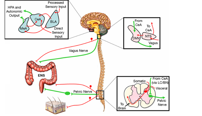
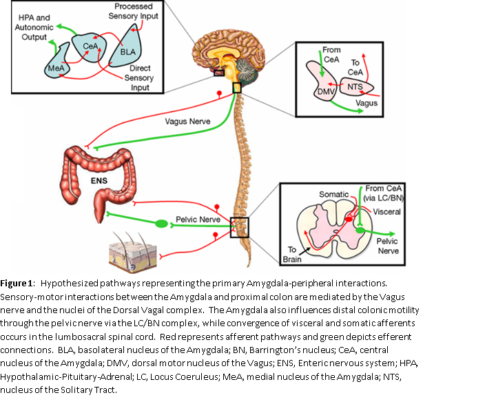
The ENS may operate at times independent of the peripheral ANS.  There is a two-way communication with the CNS and the ENS (See Figure 1).  By CNS, we are talking about the brain and the spine.  Figure 1 represents part of the “brain-gut” connection.  Abdominal pain, diarrhea, constipation, nausea, vomiting, flatulence, and abdominal distention are all symptoms of IBS.  It is believed that there are nerves in the GI tract that experience hypersensitivity and could trigger changes in the brain that perceive this as discomfort.  Many individuals who do not have these symptoms do not have this hypersensitivity of nerves in the GI tract which can trigger changes in the brain.

[1] At least three months, with onset at least six months previously, of recurrent abdominal pain or discomfort associated with two or more of the following:  1) Improvement with defecation, 2) Onset associated with a change in frequency of stool, or 3) Onset associated with a change in form (appearance) of stool.  Discomfort means an uncomfortable sensation not described as pain.

Serotonin is a major neurotransmitter (a chemical that helps nerves communicate with each other and with the organs they control) in the gut as well as in the brain. In fact, it is believed that there is more Serotonin in the gut than in the brain. It appears that Serotonin does play a part in the Brain-Gut connection. Patients with diarrhea disorders have higher levels of serotonin in their blood following a meal, and those who have chronic constipation have lower levels of serotonin than normal in the blood after meals. This has led to the development of pharmacology which can affect various receptors in the GI tract which affects Serotonin. Note, since these receptors are more specific for the gut than anywhere else in the body, this Serotonin-based sub-system is what helps to differentiate the ENS from the Parasympathetic portion of the ANS.

Serotonin receptors 5-HT3 and 5-HT4 are targeted in treatment of IBS. For example, blockade of 5-HT3 can control diarrhea and stimulation of 5-HT4 can be used to treat chronic constipation. An example of a drug that is a 5-HT3 blocker is Lotronex, which is used for the treatment of diarrhea. 5-HT3 blockers are also useful in nausea. Zofran is an example of a 5-HT3 anti-nausea medication. Zelnorm is an example of a drug that is used to stimulate the 5-HT4 receptors and can increase motility in the GI tract and improve chronic constipation states.

Newer research now is looking at Serotonin Reuptake Transporters (SERTs) which are responsible for removing Serotonin when it is released. There are reflexes in the GI tract which can stimulate diarrhea, which can be trained through relaxation techniques to operate to the patients benefit. There is a Gastrocolic Reflex which causes the colon to contract after eating large meals or fatty foods. If a patient is constipated often, it is advantageous to eat a large fatty meal or a large meal. If the patient has significant diarrhea, it is often better to eat smaller meals at more frequent intervals to not over-activate the Gastrocolic Reflexes. The gut also has its own microbiome or bacterial inhabitance. These are known as gut microbes. These gut microbes can influence and perhaps communicate with the CNS. This is a type of symbiotic relationship between us and the bacteria in our environment. These bacteria communicate through several interacting channels involving nerves, hormones and immune signaling mechanisms. This helps maintain the Brain-Gut-microbiome access. This is a bidirectional interaction from the brain to the gut involving the microbiome. Alterations in this circuit can cause IBS and other functional GI disorders.

There is current evidence that the modulation of the CNS by the microbiome occurs primarily through neuroimmune and neuroendocrine mechanisms often involving the Vagus Nerve, which is the largest nerve in the body and is the majority of the Parasympathetic nervous system. There are molecules derived from the microbes which include short-chain fatty acids, secondary bile acids and other metabolites which participate in the communication and propagation of signals through various cells.

The Blood-Brain Barrier regulates molecule traffic between the circulatory system and cerebral spinal fluid. The Blood-Brain-Barrier separates the brain from the rest of the body to protect the electrical activities that must be kept very specific and very local to the individual cells of the brain. To this end, the Blood-Brain-Barrier has very tight junctions that prevent even the diffusion of water and salts into the brain. It has been discovered that gut microbiota can change the expression of tight junction proteins, causing the junctions to become even tighter and, thereby, decrease blood-brain barrier permeability. There is communication from the brain to the gut microbiota, and it is believed that social stressors can also reduce the relative proportions of bacteria in the GI tract. Both branches of the ANS regulate gut function including regional motilities (primarily a Parasympathetic function), secreting gastric acid by carbonate mucous and other Gastric secretions (like insulin, a Sympathetic function), antimicrobials, intestinal permeability and the immuno-response from the GI mucosa. Autonomic-induced changes in gut physiology affect the microbiota that inhabit the GI tract and affect its composition and their function. Intestinal transit time, overgrowth of bacteria and other factors can affect GI motility.

Stress can cause disruption of the epithelial barrier, the so called the “Leaky Gut,” and can cause the transportation of, or “leaking out” of, gut microbes or microbe-associated molecules into the blood stream. This can cause a proinflammatory state. Excess catecholamines can affect the mucous lining of the intestinal tract. This mucous lining is protective. Inflammation of the mucous lining can occur in stress and could change the microbiota composition.

Many functional intestinal disorders have reported significant microbial shifts in the GI tract. There may be various subgroups of patients with IBS who meet Rome criteria (see Footnote 1) based on different gut microbial populations. One can do analysis of fecal levels to assess the composition of the inhabitants of a microbial community. Stress alters the ANS modulation of the gut which then can affect the composition of the micro bacteria. Specifically, stress is mediated through the Sympathetic nervous system. As an example, Lactobacilli is a natural inhabitant of the human Gut that natural protects us against invasions of harmful bacteria. A reduction in Lactobacilli has been seen during stress. Stress causes increased autonomic-Sympathetic activation.

Therefore, one can see that communication between the brain and the gut involves the microbes that inhabit the gut and the composition of these microbes can change, and are affected by Sympathetic over-activation such as stress, high blood pressure, Anxiety, poor quality sleep, cardiovascular and respiratory diseases, lack of exercise, poor diet (lack of proper nutrients), etc.; and Parasympathetic over-activation such as depression, fatigue, and trauma (mental or physical); and many other factors that are altered by abnormal changes in the Parasympathetic and Sympathetic (P&S) nervous systems.

IBS is a disorder which has been very confusing, and there has been debate for years of whether it is organic (physiologic) or psychologic in origin. It is the most common GI disorder seen in the primary care physician’s office. There are three subtypes as mentioned, a diarrhea predominant, constipation predominant and a mixed diarrhea and constipation predominant. Increasing evidence is now giving support to the theory of dysregulation within the Brain-Gut axis, which we have just discussed. Also, by studying IBS we are learning more about the so-called “second brain” or “Gut-Brain” in the intestines, which is the Enteric autonomic nervous system. The altered bowel function pain, the abdominal pain, and the hypersensitivity that is seen in IBS results from disturbances in the interaction among the gut, the brain and the P&S nervous system.

Not enough has been studied or focused on the ENS. The ENS is an extensive network of neurons supported by enteric glia which are similar to Astrocytes in the brain. Glia are cells that wrap around the nerve cell projections called dendrites that connect the nerve cell bodies. Nerve cell bodies are the decision points like little micro-processors, connected to each other and to the organs by wires, the axons. These wires are wrapped in the glial cells, of which Astrocytes are one example, like the plastic coating on wires. They protect the wire inside and to help in conducting the signal down the wire. Again, like the brain, enteric glia may also enclose large bundles of enteric axons which are the portions of the neuron that transmit electric impulses. Interestingly, the ENS contains about 100,000, 000 nerve cells which approximately equals the number in the spinal cord. Therefore, this is a very complex neural organ.

The ENS comprises two large networks of autonomic nerves known as plexuses (see Figure 1). One plexus, the Myenteric Plexus, connects to and controls or coordinates the muscles of the intestine. It is responsible for motility and is the motor plexus conducting signals from the CNS to the ENS and within the ENS. There are muscles that wrap around the circumference of the intestines to (among other actions) churn the food in the small intestines and to squeeze out water in the large intestines. There are also muscles that run along the length of the intestines to move the contents from one end to the other like a conveyor belt. Among other things the signals from these longitudinal muscles (the “conveyor belt” muscles) help to empty the bowels as more food is digested, so wastes so not back up. This “conveyor belt” example demonstrates the interaction between the Myenteric Plexus and the second plexus referenced, the Submucosal Plexus.  The combined action of the two sets of intestinal wall muscles is called “peristalsis.”

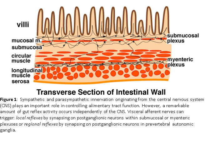
The Submucosal Plexus connects the ENS to the CNS as well as connects within the ENS.  From the simple “conveyor belt” example demonstrates these connections.  Within the ENS there are “reflexes” that work to move the contents of the intestines along in the proper direction.  They are called reflexes because they are not initiated from within the brain.  They function automatically.  However, the CNS, including the brain, needs to “know” about this motion, if for nothing else to make sure you go to the bathroom in time.  The Submucosal Plexus is the sensory plexus.  It conducts signals from sensory cells just under the mucosa of the intestine to the rest of the ENS, including the Myenteric Plexus, and to the CNS.  Among other functions, the signals from the Submucosal Plexus stimulate luminal secretions t just the right time and in just the right amount.

Signals from the brain to the gut are important for regulating digestive function and the various reflexes that occur in the GI tract and the effects of mood or psychological stress on the GI tract.  The ENS is at times autonomous and can control peristalsis and motility in the GI tract and secretion within the GI tract lumen independent of the central nervous system, but we should not rule out the central nervous system can still modulate the ENS.  For example, the “butterflies” in our stomach that we feel when nervous (like before a public speaking event or a musical or theatrical performance or before doing something very important to you) is a result of the effect that the CNS has on the ENS.  This is a base reflex.  The feeling in the stomach is designed to trigger an emptying of the bowels.  This is to “lighten the load” in case you need to run away.

Again, the most important signaling molecule involved in the peristalsis reflex, or the muscle contraction motility of the GI tract is serotonin (5-HT).  Almost all of the serotonin in the body is found in the GI tract.  We have already discussed two serotonergic receptors that are targets of pharmacologic therapy, 5-HT3 and 5-HT4.  The 5-HT3 receptors send signals indicating pain, nausea and other unpleasant sensations to the CNS.  These 5-HT receptors are involved in GI, motor and secretory functions.  The 5-HT4 receptors are help to promote peristalsis and chloride secretion and improve stool frequency and reduce bloating.

The agents that activate these selective 5-HT4 agonists (stimulants) have been beneficial in IBS with constipation in women.  In patients with diarrhea, the 5-HT3 antagonists (blockers) are helpful in controlling diarrhea IBS patients with diarrhea.  This is a complicated area but has led to development of pharmacology which can activate (stimulate) or block some of these Serotonin receptors and be beneficial in reducing symptoms of IBS of all three variants, including pain and bloating.  In IBS with constipation patients, we favor starting with Psyllium and MiraLAX and adding, if need be, Lubiprostone or Linaclotide to better improve the motility, and at times, if needed, may even add Desipramine.  Recently Motegrity (Prucalopride) was approved for IBS with constipation.  It is a Serotonin agonist.  In IBS with diarrhea patients, we may use low dose Loperamide or even Rifaximin, a non-absorbable antibiotic, and if need be add very low dose tricyclic antidepressants (TCAs) which can control the neural hypersensitivity of abdominal discomfort and diarrhea.  TCAs are antidepressants at clinical doses (e.g., around 100 mg per day), but function as anticholinergics at low doses (of no more than 10 mg per day, and at the lower dose, the known side effects of antidepressants are present, including suicide risk).  We also favor other antidepressants such as Selective Norepinephrine Reuptake Inhibitors (SNRIs) over Selective Serotonin Reuptake Inhibitors (SSRIs).  For more advanced therapy, sometimes a drug called Alosetron is recommended.  Alosetron works when a 5-HT4 mechanism is involved.  Gastric bloating, including that associated with constipation, is difficult to treat in IBS.  Low doses of high fermenting sugars and empiric Rifaximin have been advocated at times.

In summary, the pathophysiology of IBS is very complex.  There is abundant data that supports visceral hypersensitivity alterations in the gut microbiome, intestinal permeability (“Leaky Gut Syndrome”), gut immune function and motility changes, brain-gut interactions, and psychosocial stress all may contribute to the development of IBS.  Pain after eating meals reflects altered motility and may be an expression of a heightened Gastro-Colic Reflex.  Increased permeability, which may be a mechanism in development of IBS will be discussed later under Leaky Bowel Syndrome where disrupted tight junctions between cells in the GI tract lead to increased permeability of toxins and bacterial products into the blood stream.  This exposes the enteric nerves or nerves in the GI cells to stimuli which can cause the pain and discomfort in visceral hypersensitivity.  After viral infections or gastroenteritis infections, IBS may actually emerge.  Many feel it is the result of abnormalities that occur in the tight junctions between cells in the GI tract causing a “Leaky Gut.”  Altered gut microbiota, as discussed earlier, may be associated with gut immune function and altered gut motility and can also lead to hypersensitivity in patients with IBS.

Again we should not discount psychosocial factors, as it has been shown that early life stress may cause development of exaggerated pain perception in patients with IBS, and we will often recommend stress relaxation techniques such as yoga and meditation or prayer to these patients.

Ultimately, IBS is a diagnosis of exclusion.  While one has to fulfill the Rome criteria, or its definition, one really needs to exclude other more serious disorders and oftentimes a Gastroenterologist will exhaust many invasive and noninvasive tests in the process to exclude tumors, colitis, and infections.  Note, the tests that take pictures of videos of the GI tract (Endoscopes, Colonoscopy, and the Smart Pill camera) assess the anatomy of the GI tract to look for blockages, etc.  Motility disorders, such as IBS, also require physiologic tests to determine transit time and efficacy of peristalsis.

The first step in treating IBS in general, whether constipation or diarrhea predominates, is using a Low-FODMAP diet, which is a diet low in fermentable sugars (oligosaccharides and disaccharides).  With this diet improvement in bloating, abdominal pain, and flatulence may improve, as well as the altered bowel movements.  A dedicated diet needs to be adhered to in these cases.  Gluten-free diets may also be effective, but there is debate over this issue.  Soluble fiber seems to do a better job in the constipation-predominate IBS and antispasmodics, which include Dicyclomine and Hyoscyamine, and even Peppermint Oil, may be useful to relieve the abdominal pain and cramping.   Antispasmodics, over some anticholinergics, may cause constipation and are preferably used in IBS with diarrhea.  Antidepressants, especially tricyclics, may be useful in low dose.  We already discussed IBS constipation specific medicines, Lubiprostone (Amitiza) and Linaclotide (Linzess).  These are useful in constipation-predominant IBS.  In diarrhea-predominant IBS, antidiarrheal agents may be used, bile acids such as Colestipol or Cholestyramine may be used, and even Welchol (which is used to lower cholesterol and also used to lower sugar in diabetes)  Tricyclic antidepressants are more useful in diarrhea type of IBS including Amitriptyline, Nortriptyline, and Desipramine, although we will use Desipramine in constipation-predominant at times.  Alosetron (Lotronex) blocks the extra serotonin 5-HT3 on the nerves in the GI tract and will slow the motility of the GI tract and improve diarrhea.  It will also reduce pain and distention as well as flatus.  However, side-effects such as Ischemic Colitis, perforation and death have been noted and therefore a GI specialist who is experienced in using it should prescribe the medication, in women specifically who have diarrhea predominant IBS.

Rifaximin (Xifaxan) is a nonabsorbable antibiotic, which has been studied in an IBS diarrhea-type in randomized trials and can be used in retreatment when flare-ups occur.  However, it has side-effects of flatulence and abdominal pain and nausea, which may limit its use; also it is extremely expensive.  One needs to seek more attention with the Gastroenterologist if they have uncontrolled IBS with constipation or diarrhea.  Recently, an approved medication for diarrhea, Viberzi, is an antidiarrheal agent which operates through opiate receptors and may be useful.

Lastly, since there is a psychosocial component and a stress component which adversely affect the ANS and can aversely affect the microbiota in IBS patient, psychological interventions may be useful including cognitive behavior and relaxation techniques, such as yoga and meditation and prayer.  Exercise and stress modulation are extremely important in treating patients.  Acupuncture may also be helpful in some people, but there have been conflicting results.  These activities help to normalize Parasympathetic activity which is a key factor.

There are two possible Parasympathetic effects:  Parasympathetic Insufficiency and Parasympathetic Excess.  Since the Parasympathetics, through the Vagus Nerve and the ENS, have the primary control of the GI tract, we should consider the interaction between the Parasympathetics and the GI tract for a moment.  Parasympathetic Insufficiency indicates abnormally low Parasympathetic activity, which may cause abnormally slow GI motility, which may involve Gastroparesis and constipation.  Parasympathetic Excess indicates abnormally high Parasympathetic activity, which may cause abnormally fast GI motility, which may involve diarrhea.  Both may also cause Sympathetic Excess.

Consider a car, with brakes and an accelerator.  The brakes are like the Parasympathetic nervous system and the accelerator is like the Sympathetic nervous system.  In cases of Parasympathetic Insufficiency, motility is very low because there is no Parasympathetic activity to drive it, but with weak or no brakes, the accelerator (the Sympathetics) may be excessive in these cases as well, because there is no way to slow them.  Therefore, Sympathetic symptoms including amplified pain, too much insulin, anxiety, and other stress symptoms predominate.  With Parasympathetic Excess, it is like driving a car with a foot on the brakes.  A foot on the brakes cases all accelerations to also be excessive just to get to normal speed; the engine is being over-revved to over-come the brakes.  This is also Sympathetic Excess and may cause the same symptoms.  The difference is whether there is motility or not.  In cases of Parasympathetic Excess, the fact that at different times (changes in hormone levels, stress levels, emotional levels, exercise or diet, etc.) IBS may be associated with constipation or with diarrhea may be more understandable.  When Parasympathetic Excess predominates, diarrhea is more likely.  When the reactive Sympathetic Excess predominates, constipation is more likely.  In both cases, getting the foot off the brakes helps to relieve the IBS and both diarrhea and constipation.

We should not discount the stress effect.  During periods of increased Anxiety, hormones, such as Cortisol, Adrenalin and Serotonin are released by the brain, and this will raise the amount of Serotonin in your GI tract and cause stomach spasms to occur.  If the spasms occur throughout your entire colon, an individual can get diarrhea because of a hypercontraction issue, but if the spasms are located to one area of the colon such as the sigmoid colon, one area which is extremely susceptible and which is almost considered an “Achilles heel,” of the GI tract, digestion may actually be curtailed and stopped and constipation may result.  Therefore, a spasm which occurs with stress can be focal which can cause constipation or diffuse which can cause diarrhea and is usually mediated by high quantities of Serotonin in your gut after first being released from the brain.

Also, cortisol and adrenaline released from the brain can be adversely effective in times of stress.  The stress, as mentioned, causes bacteria in the GI tract to become imbalanced.  The term for this is Dysbiosis and may specifically contribute to IBS-related constipation.  In more serious GI conditions, known as inflammatory bowel disease (IBD), stress can cause a flare up in these disorders.  It is believed that chronic stress (a Sympathetic response) and depression (a Parasympathetic response), both occurring together as with Parasympathetic Excess, appear to increase inflammation (a Sympathetic Excess) which may set off the flares in IBD patients.  Just as Anxiety (another Sympathetic Excess) may cause worsening of IBS or IBD flare-ups. Having these diseases and the comorbidities and symptoms associated with them may also cause Anxiety or more Anxiety, and this may cause a vicious cycle.

The cause of IBS for many years was thought to be largely psychogenic.  However, now we know it is multifactorial.  The identification of microbes which inhabit the GI tract and an imbalance which can occur with autonomic (Parasympathetic or Sympathetic) dysfunction and stress has been recognized to possibly cause gut Dysbiosis.  Recently, a disorder known as Small Intestinal Bacterial Overgrowth has been noted to be associated with, or possibly cause, IBS symptoms.  The fact that some probiotics and some absorbable and non-absorbable antibiotics may help improve symptoms in patients with IBS suggests that IBS may not originate in the brain but rather IBS may predominately originate in the GI tract (the ENS or the “Gut-Brain”) and supports a microbe altering basis for IBS.

SIBO is present when there is an increase in bacteria equal to, or greater than, 105 colony-forming units per mL in an upper gut aspirate test.  These patients also experience abdominal pain, discomfort, bloating, flatulence, loose stools and may even have constipation, if one of the gasses secreted is methane and is in high quantities.  It was once thought that SIBO only occurred in patients who have intestinal anatomical obstructions or malformations, but it is now realized that it may occur in the absence of anatomical factors predisposing to it.  The gold standard for diagnosis is an aspirate from the small bowel and quantitating the amount of bacteria present.  There are breath tests available; however, these can give false positives.  These breath tests measure hydrogen, methane and hydrogen sulfide.  They are simpler than doing a direct culture in the GI tract.  However, as noted, there may be false-positives and the correlation with the gold standard stool cultures in the small bowel may not be precise.  However, it is easy to follow patients with a breath test as these may be reproduced when therapy is given.  For example, when an individual undergoes therapy for SIBO with antibiotics, one could measure a breath test and as it improves oftentimes the symptoms are improving and one knows that the SIBO is being treated appropriately.  The two most commonly used substrates for testing for SIBO are Glucose and Lactulose.  Lactulose, however, can cause diarrhea by itself and can hasten intestinal transit time; thereby, giving a false-positive breath test.

Testing for methane has become extremely important especially in identifying people who have chronic constipation disorders.  The methane itself may slow intestinal transit and cause significant constipation.  It is believed that microorganisms called Archaea, which are predominately found in the colon, may start populating the small intestine when there is significant methane gas present.  The methane gas is produced by Archaea organisms.  One can measure the bacteria Methanobrevibacter Smithii and correlate the levels of this bacteria with the degree of constipation individuals may have as this bacteria predominately secretes methane.  The most common symptom with SIBO is bloating.  More bacteria in the small bowel theoretically may cause a greater capacity for gas production and more abdominal distention and cramps.  Flatulence and belching may become prominent.

There are many conditions associated with SIBO.  These include motility disorders, such as gastroparesis, IBS, pseudo-obstruction, and constipation disorders known as colonic inertia, and mechanical abnormalities, such as adhesions and strictures, often seen with inflammatory bowel diseases, such as Crohn’s disease, bowel obstruction, polyps and tumors.  Also Diabetes and Achlorhydria are sometimes associated with SIBO.  Oftentimes, this is associated with Proton Pump Inhibitors (PPIs).  There are immune mechanisms which may predispose to SIBO including deficiencies of IgA, collagen vascular diseases such as scleroderma and lupus, immunoglobulin abnormalities such as common variable immunodeficiency, HIV infections, chronic pancreatitis, acute pancreatitis episodes may also predispose to SIBO, as well as Cirrhosis.  Ehlers-Danlos Syndrome has been known to predispose to SIBO.  Medications such as opiates, anti-diarrheal agents, which slow GI transit, and acid-reducing agents specifically PPIs have been associated with SIBO.  However, the most common disease associated with SIBO is IBS.  This is often seen after an acute episode of Gastroenteritis where, especially females, can develop a new-onset of IBS and SIBO concurrently.

Besides IBS, conditions that have been associated with SIBO include Inflammatory Bowel Disease, Rosacea, Dyspepsia, Restless Leg Syndrome, Small Bowel Diverticula, Pancreatitis as noted, Hypothyroidism, Parkinson Disease, Diabetes, Coronary Artery Disease, abdominal surgery such as Hysterectomy, Cholecystectomy, Gastrectomy and Colectomy.  SIBO is largely underdiagnosed.  Risk factors for SIBO need to be sought for in taking a history of a patient with abdominal symptoms to potentially diagnosis more patients who have it.  Again, low stomach acid, IBS symptoms, Celiac Disease, long-standing Crohn’s Disease, and other inflammatory bowel disease, prior bowel surgery, Diabetes Mellitus with type 1 and type 2, multiple courses of antibiotics, and organ dysfunction, such as Liver Cirrhosis, Chronic Pancreatitis or Renal Failure may predispose to SIBO along with abnormalities in the GI tract.

The goal of treating SIBO is symptom relief by eradicating any of the significant overgrowth of bacterial in trying to bring the GI flora back to a normal balance.   Antibiotics are oftentimes the mainstay if diet is not effective.  Sometimes the bacteria may be antibiotic resistant and there may be other underlying conditions, such as dysmotility or use of drugs such as PPIs that need to be sought after and corrected.  One should treat predisposing conditions that cause SIBO.  This is especially true if it was put in remission since it can recur.  Promotility drugs and bowel preps with various types of laxatives to keep bowel motility functioning may be important in preventing the recurrence of SIBO.  It is said that 44% of patients with SIBO may experience a relapse of symptoms within nine months of initial treatment.  Avoiding medicines that delay gut transit time, improving glycemic control in diabetics, and correcting any anatomical abnormalities such as blind intestinal loops if they are present are the first line of treatment.

One should speak to their Gastroenterologist about using a prokinetic drug, even prophylactically so an occurrence does not recur.  Many of these prokinetic drugs are the same ones that are used for gastroparesis or constipation and include Cisapride, Tegaserod, Erythromycin and the newly approved drug, Prucalopride.  Some of these agents have risks and risk/benefit ratios have to be discussed with the physician.  Some experts have even used low dose antibiotics rotating them cyclically every month and use two or three antibiotics that are known to be effective for these disorders.  One should remember, however, that SIBO is not the explanation for all bloating abdominal pain or altered bowel habit.  Whether positive for culture or negative for culture, it may be just small intestinal dysbiosis or an abnormal balance or other gut pathologies which may cause SIBO or be caused by SIBO.

Also, herbal microbials have been used as a good option to treat Colonic Dysbiosis and have fewer side-effects.  The most successful antibiotic is the nonabsorbable antibiotic Rifaximin.  Also, gut-directed stress management is important.  Some patients have required a combination of Rifaximin and Neomycin, both nonabsorbable antibiotics, especially in constipation-predominant SIBO patients with methane-predominant bacterial overgrowth.  Other antibiotics used have been Ciprofloxacin, Metronidazole, Amoxicillin-Clavulanic Acid and several others.  Elemental diets have also been shown to be effective in 80% of patients with methane or hydrogen-predominant SIBO.  These diets were originally developed for short bowel syndrome but now have extended use in patients with normal bowel structure who have SIBO.  Interestingly, statins can also inhibit methane gas production directly and have been found to be useful in these patients.

Healthy exercise, proper diet and stress reduction are the most effective for long-term balance; both in the Gut Microbiome and in autonomic (P&S) balance – they go hand in hand.  Furthermore, there is often no quick-fix.  Both systems are like pendulums.  They cannot be corrected with a sledge-hammer, it has to be gentle nudges over time.  Quick fixes often push the balance too far in the opposite direction and lead to more or worsening symptoms.

Of course, with patients with SIBO, if there is an underlying disease it should be treated.  For example, a flare-up of Crohn’s Disease or Celiac Disease should be treated directly along with SIBO.  Diabetes should be treated, and avoidance of diabetic drugs known to slow the gut motility, such as Glucagon-like peptide 1-agonists should be avoided.  We have found that patients with connective tissue disease and joint hypermobility syndromes (including EDS) benefit from pro-motility drugs.  Therefore, treating SIBO and even patients with IBS who have not undergone testing for SIBO should concentrate on diets which can manipulate gut microbiota beneficially.  Vegetarian diets rich in fiber lead to higher production of short chain fatty acids which inhibit potentially invasive bacteria like E. coli.  Diets rich in complex carbohydrates favor a growth of less pathogenetic bacterium than diets rich in fat or protein.

The recent recognition that SIBO plays an important role in the pathogenesis of patients with IBS has led us away from a psychological etiology for the small bowel symptoms and disturbances.  Many patients with concomitant autonomic dysfunction also have microbe abnormalities in the GI tract.  SIBO is often associated with brain fogginess, and patients who receive different antibiotics report improvement of SIBO-related symptoms in over 70% of patients.  This is further evidence of a physiologic and potential autonomic dysfunction.  In many cases brain-fog is a function of poor brain perfusion associated with autonomic dysfunction.  However, there is no agreement as to the frequency of SIBO among IBS patients.  Some studies have shown as low as 4% and some as high as almost 80%.  It is hoped that stool sampling with various immunological techniques may become more practical and sensitive and specific than small bowel culture or oral breath tests in the future.  At the present time, Rifaximin is the best treatment for SIBO among patients with irritable bowel syndrome based on the totality of the data reviewed so far.  Rifaximin should be prescribed by a Gastroenterologist and followed carefully as recurrences may need to be retreated.

One should not forget that there are other natural treatments besides diet such as probiotics, which include lactobacillus, Bifidobacterium, Saccharomyces, and other mixed compounds.  In rare cases, SIBO may be precipitated by probiotics.  However, as mentioned, this is extremely rare.  Again, herbal supplements have also been proposed to be beneficial.

As mentioned, there is a brain-gut and microbiota miscommunication in patients with significant troubling GI symptoms.  The peripheral ANS has been evaluated in many of these patients, although we now realize that the Enteric-ANS is also functioning independently especially with the transmitter Serotonin.  If one looks at patients with IBS and constipation, one finds impaired Sympathetic activity and disturbed Parasympathetic function.  It has been postulated that a central Sympathetic influence within the brain-gut axis is probably responsible for the myoelectrical activity disturbances in IBS patients [1].  These patients have increased Insulin, Norepinephrine and Epinephrine secretion; which are all results of Sympathetic activity.

IBS is associated with behavioral factors and stress hormone pathogenesis and can increase these hormones.  These can increase insulin resistance and create Sympathetic hyperactivity or Sympathetic Excess.  Heart Rate Variability (HRV) testing has been used to demonstrate Sympathetic Excess.  P&S imbalance can be corrected with normalized ENS activity.  The high frequency component of HRV is a measure of Vagal, or Parasympathetic, tone.  The ratio of the low frequency HRV component to the high frequency component is an indicator of Sympathovagal Balance (SB, aka, P&S Balance).  A meta-analysis has shown that the high frequency or SB components of HRV are affected adversely in a significant proportion of patients with IBS.  IBS patients show higher SBs, indicative of a relatively high sympathetic tone at rest as compared with resting Parasympathetic tone.  Also, constipation-predominant IBS patients had decreased high frequency activity indicative of low Parasympathetic activity at rest.  Other studies of IBS patients have shown abnormalities in cardiac bio-reflex with ANS testing in patients who have autonomic dysregulation.

Therefore, we do recognize abnormalities in the peripheral ANS which can be identified in routine testing in the office setting and balance can be attempted pharmacologically in these areas.  However, a recent review [2] describes orthostatic intolerance and postural tachycardia and its association with GI symptoms.  The review states that only when the GI symptoms develop in the upright position and then resolve on lying flat can we say that they are causative and related directly to the orthostatic stress.  The most common symptoms associated with orthostatic intolerance, include nausea, dyspepsia, bloating and constipation, yet the majority of the subjects do not have gastroparesis.  They believe that Postural Orthostatic Tachycardia Syndrome (POTS) is comorbid with many other symptoms such as Migraine Headaches, Fibromyalgia, Chronic Fatigue Syndrome, sleep disorders, abdominal pain, and joint hypermobility (EDS), etc.  Further, it is believed that these GI symptoms are also a comorbidity affecting orthostatic intolerance symptoms in POTS patients.  They believe POTS is not the driver of the comorbidities, which means that autonomic dysfunction syndromes, such as POTS or orthostatic intolerance symptoms can coexist with GI symptoms but are separate.  Although, one cannot discount the common mechanism of brain, heart, blood vessel and brain-gut communications having at times similar mechanisms.

During tilt testing, if gastrointestinal symptoms occur, they usually include nausea and abdominal pain.  This suggests that some of these symptoms are related to orthostatic challenges but do not occur universally in all patients. These patients who do have nausea and abdominal pain with tilt usually do respond to volume expanders like fludrocortisone.  However, we recognize that treating POTS and orthostatic intolerance with autonomic agents oftentimes does not beneficially affect their GI symptoms if they are not reproduced with tilt test or assuming the upright position (e.g., standing-up).  The presence of POTS does not seem to influence the general findings of chronic overlapping pain conditions with functional GI disorders.  Our clinical observation, and the empiric observations of others, suggests that a lower vagal modulation may be associated with the more chronic pain disorder rather than what the POTS subjects have in general.  Also, these observations suggest that Vasovagal Syncope is more often co-morbid with POTS that previously accepted.  Further suggesting Vagal or Parasympathetic dysfunction is involved, including the ENS.  Again imbalance in the ENS, Serotonin transmission, and the gut-brain microbiota circuit miscommunications are implicated in IBS.

Therefore, as an autonomic physician, when I see patients with orthostatic intolerance and POTS, even if their GI symptoms may not be directly related, it is important to recognize and understand that the nausea, abdominal pain, constipation, bloating and diarrhea must be managed concomitantly with the P&S dysfunction(s), even though the drug treatments and lifestyle treatments may be different.  Interestingly, an altered bowel pattern has been reported in 70% of POTS patients.  Diarrhea and constipation, however, are not triggered usually by upright position and tilt.  Therefore, they should be treated separately.

In regard to migraines, they are often associated with nausea.  This also suggests the involvement of the ANS, specifically the Vagus nerve of the Parasympathetic nervous system.  Vagal symptoms are well known in the development of migraine with nausea.  The nausea may also be due to autonomic dysfunction in the form of orthostatic challenge, and therefore, one needs to differentiate this.  In fact the migraine may also be due to autonomic dysfunction in the form of poor brain perfusion (blood flow to the brain).  If the nausea is not relegated to orthostatic challenge and there is no delayed gastric emptying, patients with migraine and nausea can be treated with Cyproheptadine or tricyclic antidepressants or Topiramate.

Dyspepsia is another disorder in which oftentimes a clear-cut mechanism or etiology cannot be found and slow gastric emptying is not present.  These patients are oftentimes treated with prokinetic agents like Erythromycin, cholinesterase inhibitors, Buspirone (relaxes the gastric fungus), as well herbal products and tricyclics at low dose, which are better tolerated than SSRIs.  One needs, of course, to exclude H. Pylori in these patients.  When treating dyspepsia, one starts with H2 blocker or PPPIs to decrease the acid and then can go through the other medicines we have just discussed to see if there is empiric benefit.  One must first demonstrate there is a normal upper endoscopy and no H. pylori infection in people who present with dyspepsia.  Occasionally, cognitive behavioral therapy and peppermint oil may be used, or occasionally one needs to go to prokinetic agents if tricyclics and SSRIs/SNRIs are not beneficial.

There is a case report of a special treatment of POTS with mast cell activation syndrome using Naltrexone, Immunoglobulins and antibiotic treatment.  These authors discussed Intravenous Immune Globulin (IVIG) as an emerging promising therapy for POTS.  This is an immunomodulating agent and response to this suggests that some of the autonomic imbalance seen in some POTS patients may be due to active autoimmune muscarinic antibodies against acetylcholine.  Many mast cell activation syndrome patients do have GI abnormalities and we should exclude SIBO and IBS in these patients.  However, we are not anxious to use immunomodulating agents without more concrete evidence, such as autoantibodies, paraneoplastic antibodies, and so forth, which we can test for.  Oftentimes, a consultation with an immunologist and a rheumatologist can be beneficial.  There is a link between the autonomics and the immune system, specifically the Parasympathetics control and coordinate the immune system, including Sympathetically mediated histaminergic responses as effected by mast cells.

Vagal, or Parasympathetic, dysfunction with SIBO has been recognized.  In a study out of Mt. Sinai Hospital in New York, a subset of patients with chronic inflammation with HIV infection had abnormalities when components of the Vagal nerve were tested.  These abnormalities correlated with changes in immune function and GI function in well treated patients with HIV infection.  It was postulated that possibly enhancing Vagal function could help benefit HIV patients in the future.  The authors emphasized that a function of the peripheral ANS is promotion of GI motility by cholinergic fibers of the Vagus nerve.  A potential consequence of Vagal nerve fiber losses slowed motility particularly in the stomach and proximal small intestine with a Vagal nerve context with Enteric neurons.  With this slowed motility there is propensity for SIBO.  This can promote bacterial translocation, which is what we see in the increased gut permeability syndrome and drive inflammation.

At the other end of the spectrum, cholinergic stimulation, such as provided by the Vagus nerve, has also been shown to modulate GI mucosal inflammation and increase mucosal populations of CD4 cells.  CD4 cells are white blood cells that play an important role in the immune system. They provide your body’s natural defense against pathogens, infections and illnesses.  CD4 cells are sometimes also called T-cells, T-lymphocytes, or helper cells.  Your CD4 cell count gives an indication of the health of your immune system.  Independent of the effects on GI motility, Vagal dysfunction could also contribute to chronic inflammation by direct effects on the immune system.  These investigators tested patients with sudomotor testing, beat-to-beat blood pressure with tilt and conventional HRV-derived parameters with standard Ewing maneuvers such as Valsalva.  They developed a Composite Autonomic Severity Score (CASS) which included sudomotor, Vagal and adrenergic sub-scores.  They showed that Vagal dysfunction was associated with slowed gastric emptying and SIBO.  They also correlated this with elevated levels of IL-6 and other inflammation markers.  They postulated that it is in the GI mechanisms that Vagal dysfunction may be linked to immune dysfunction in HIV patients, and also Vagal dysfunction is linked to chronic inflammation.

Impaired intestinal barrier integrity in the colon of patients with IBS has been demonstrated in prior studies.  This involves soluble mediators.  These most likely also contributed to chronic inflammation in IBS patients.  Again, studies on IBS patients have shown abnormal Vagal dysfunction.  Increased GI permeability or “Leaky Gut Syndrome” is a real condition.  Simple tests such as Lactulose-Mannitol Intestinal Permeability tests can be done especially when assessing Celiac Disease patients for increased permeability.  Other more sophisticated tests such as colonic biopsies looking for mRNA expression in tight junction proteins as has been done with IBS patients to demonstrate increased intestinal permeability is a more elaborate test.  There are many tests to assess the intestinal barrier.  They have all shown that abnormalities can occur in patients with various GI disorders, such as SIBO and IBS.  They have also shown accompanying inflammation and abnormal Vagal tone can be associated with it.

While further research is needed, we strongly advocate in our patients balancing any abnormalities in the general peripheral autonomic (or P&S) nervous system that we encounter especially if there is accompanying orthostatic intolerance or other postural disorders.  We advocate exercise, a proper anti-inflammatory diet, and antioxidant supplements along with nitric oxide boost of beetroot.  Both stress reduction at the cellular level (known as oxidative stress) and at the whole body level (known as Psychosocial stress) is also a significant part of treating any patients who need to have their P&S nervous systems in balance and who need to have their GI system balanced to function better.


REFERENCES

[1] Mazur M, Furgała A, Jabłoński K, Mach T, and Thor P.  Autonomic nervous system activity in constipation-predominant irritable bowel syndrome patients.  Med Sci Monit. 2012;18(8):CR493–CR499. doi:10.12659/msm.883269.

[2] Chelimsky G, Chelimsky T.  The gastrointestinal symptoms present in patients with postural tachycardia syndrome: A review of the literature and overview of treatment.  Auton Neurosci. 2018 Dec;215:70-77. doi: 10.1016/j.autneu.2018.09.003. Epub 2018 Sep 8.

Read More