More Than Sick of Salt

All Posts in Category: parasympathetic nervous system

Coronavirus Induces Oxidative Stress Leading to Autonomic Dysfunction – Part 2

Click here to download this post

Notes: This is the second in a series of 5 blog posts about COVID-19 and Autonomic Dysfunction.  This a pre-publication release that will be featured in a major medical journal.

Coronavirus Induces Oxidative Stress Leading to Autonomic Dysfunction Often With Delayed Symptom Onset

Heather L. Bloom, MD1 and Joseph Colombo, PhD, DNM, DHS2

  1. Electrophysiology, Atlanta Veterans Affairs Medical Center and Emory University Medical School, Atlanta, GA 
  2. Parasympathetic & Sympathetic Nervous System Consultant, Franklin Cardiovascular Associates, PA & Autonomic Dysfunction and POTS Center, Sewell, NJ, and Senior Medical Director & CTO, Physio PS, Inc., Atlanta, GA, dovetech@erols.com

Correspondence should be addressed to Dr. Colombo, dovetech@erols.com

INTRODUCTION

It is generally well known that many chronic and serious pathologies cause an over-production of oxidants, including Reactive Oxygen Species (ROS), Reactive Nitrogen Species (RNS), and many other oxidative molecules. 

What may not be as well-known is the fact that severe acute conditions may also cause an over-production of oxidants.  A recent published review [[i]] highlighted this in COVID-19 patients. 

Many of the other pathogens that cause severe acute diseases are also implicated, including Influenza (like COVID19, a SARS virus) and many other viruses, bacteria like the Borrelia bacterium that causes Lyme Disease, severe physical or physiological stresses or traumas like that which triggers what is known as Fibromyalgia; as well as severe exposures to cold, heat, chemicals, etc., and severe mental or emotional traumas (e.g., PTSD); to name a few.

An over-production of oxidants is known as Oxidative Stress.  While some level of oxidants are necessary for the Immune system as a first-line defense against pathogens, for programmed cell death and other general cellular house-keeping activities, too many oxidants lead to cell and organelle damage, including, damage to Mitochondria. 

The cardiovascular and the nervous systems have the highest numbers of Mitochondria per cell and are therefore more susceptible to Oxidative Stress. 

As the cardiovascular tissue and the Parasympathetic and Sympathetic (P&S) branches of the autonomic nervous systems (ANS) become disordered, P&S dysfunction accelerates cardiovascular disorder and a downward spiral begins; often long before recognized disease symptoms present. 

Further, in addition to collecting oxidants for beneficial use, the Immune system is primarily responsible for balancing the oxidants and antioxidants in the system. With P&S dysfunction this balancing process becomes less effective.

Oxidative Stress-induced P&S Dysfunction may be associated with a huge constellation of symptoms and conditions including  Lightheadedness, fatigue, wild fluctuations in blood pressure, blood glucose, hormone levels, and weight; as well as difficult to describe pain syndromes (including complex regional pain syndromes, or CRPS), excessive symptoms of palpitations without clinical correlation to definitive pathology or seizures, temperature dysregulation (to heat and/or cold and sweat responses), and symptoms of depression and anxiety, ADD/ADHD, exercise intolerance, sex dysfunction, sleep or GI disturbance, cognitive dysfunction or “brain fog”, or frequent headache or migraine.

Given the current high volume of the Severe Acute Respiratory Syndrome Coronavirus 2 (SARS-CoV-2, or Coronavirus Disease 2019, or COVID-19 virus, or COVID), it will be the exemplar severe acute pathology used as a model of other severe acute pathologies. 

In patients that recover from COVID, the basic model is: 

1) COVID causes Oxidative Stress in most patients who recover;

2) The severity of the resulting Oxidative Stress is debilitating to a sub-population of the patients affected (perhaps 15%, in the case of COVID, [personal clinical observations]);

3) Oxidative Stress damages cell membranes, DNA, and (especially) Mitochondria;

4) as the cells that utilize the most energy (ATP) in the body, nerve and cardiovascular cells are the most susceptible to Oxidative Stress damaged Mitochondrial dysfunction;

5) Mitochondrial dysfunction in the P&S nerve cells themselves and the mitochondrial damaged cardiovascular cells, both cause changes in P&S that manifest three to six months after relief of the initiating pathology (COVID);

6) due to this delay, and the normalcy of the interim, the resulting P&S Dysfunction is not associated with COVID,

7) the resulting P&S Dysfunction causes many symptoms the most complained about is lightheadedness and persistent fatigue that is not treatable by standard therapies.

Both Oxidative Stress and P&S imbalance are treatable [[ii]], depending on individual history, 9) rebalancing oxidation and P&S leads to improved outcomes including quality of life (i.e., fatigue is relieved) and improved productivity [[iii]].

The main clinical dilemma is that the connection between COVID and P&S Dysfunction is not obvious.  The symptoms of P&S Dysfunction, presenting three to six months after the disease is relieved and apparent normal function is returned, are interpreted as a new condition and misinterpreted as not a continuation of the previous condition. 

This causes three problems.  First, since only the symptoms are being treated, the therapy plan is often confounded due to conflicting dysfunctions.  For example, fatigue is often accompanied by lightheadedness or dizziness, anxiety, depression, sleep difficulties, and loss of productivity. 

Treating all of these symptoms individually involves competing agents.  Furthermore, and the second problem, since what is being treated are symptoms and not the underlying cause (Oxidative Stress and P&S imbalance), therapies are usually titrated to higher doses; and yet, the patients still do not respond as expected. 

Moreover, if and when P&S Dysfunction is suspected, the high doses of these medications often leave the patient sensitized to these medications.  This sensitization precludes their use at the very low levels needed for balancing the P&S nervous systems. 

All of these treatment issues can leave the Physician thinking that the patient is non-compliant or psychosomatic, which often leads to a psychology referral.  This can lead to the breakdown of the physician-patient relationship since the patient is sure that the symptoms are real and not in her or his head.

As suggested by the title of the recently published article [i], a simple P&S assessment may be made in the clinic to identify any P&S imbalance.  Relieving the P&S imbalance, which often involves Antioxidants [ii], and thereby restoring P&S and oxidant balance, relieves or prevents the symptoms of P&S imbalance post-COVID; thereby, minimizing any further reductions in quality of life and losses in productivity.

P&S testing is not ANS testing.  Most ANS test only test total autonomic function and force assumption and approximation to theorize P&S activity.  There is only one P&S test that provides simultaneous, independent measures of P&S activity.  What is expected from P&S testing is one or more of four possible P&S Dysfunctions that underlie the Dysautonomia typically associated with Oxidative Stress.


REFERENCES

_________________

[1] Murray GL.  COVID-19 cardiac complications: Is an easy, safe treatment strategy right under our noses?  J Cardiovasc Dis Diag. 2020; 8:5.  doi: 10.37421/jcdd.2020.8.415.

2 DePace NL, Colombo J.  Autonomic and Mitochondrial Dysfunction in Clinical Diseases:  Diagnostic, Prevention, and Therapy.  Springer Science + Business Media, New York, NY, 2019.

3 Acosta C, DePace NL, DePace NL, Kaczmarski K, Pinales JM, and Colombo J.  Antioxidants effect changes in systemic parasympathetic and sympathetic nervous system responses and improve outcomes.  Cardio Open. 2020; 5(1): 26-36.  doi:  10.33140/COA.05.01.04

4 Colombo J, Arora RR, DePace NL, Vinik AI.  Clinical Autonomic Dysfunction:  Measurement, Indications, Therapies, and Outcomes.  Springer Science + Business Media, New York, NY, 2014.

5 Vinik A, Ziegler D.  Diabetic cardiovascular autonomic neuropathy.  Circulation. 2007; 115: 387-397.

6 Vinik AI, Maser RE, Nakave AA.  Diabetic cardiovascular autonomic nerve dysfunction.  US Endocrine Disease.  2007; Dec: 2-9.

7 Malik, M.  The Task Force of the European Society of Cardiology and the North American Society of Pacing and Electrophysiology. Heart rate variability, standards of measurement, physiological interpretation, and clinical use.  Circulation. 1996; 93:1043-1065.

8 Malik, M. and the Task Force of the European Society of Cardiology and the North American Society of Pacing and Electrophysiology. Heart rate variability, standards of measurement, physiological interpretation, and clinical use.  European Heart Journal. 1996, 17: 354-381.

9 Akselrod S, Oz O, Greenberg M, Keselbrener L.  Autonomic response to change of posture among normal and mild-hypertensive adults: investigation by time-dependent spectral analysis.  J Auton Nerv Syst. 1997 May 12;64(1):33-43.

10 Piña IL, Di Palo KE, Ventura HO.  Psychopharmacology and Cardiovascular Disease.  JACC. 2018; 71(20): 2346-2359.

11 Arora RR, Bulgarelli RJ, Ghosh-Dastidar S, Colombo J.  Autonomic mechanisms and therapeutic implications of postural diabetic cardiovascular abnormalities.  J Diabetes Science and Technology.  2008; 2(4): 568-71.

12 DePace NL, Vinik AI, Acosta C and Colombo J.  Oral vasoactive medications:  A Review of Midodrine, Droxidopa, and Pseudoephedrine as Applied to Orthostatic Dysfunction.  NEJM.  2020.  Submitted.

13 Vinik AI, Bloom HL, Colombo J.  Differential effects of adrenergic antagonists (carvedilol vs. metoprolol) on parasympathetic and sympathetic activity:  A comparison of measures.  Heart International. Heart Int. 2014; 9(1): 7-14; DOI: 10.5301/HEART.2014.12495.

14 Bloom HL, Vinik AI, Colombo J.  Differential effects of adrenergic antagonists (carvedilol vs. metoprolol) on parasympathetic and sympathetic activity:  A comparison of clinical results.  Heart Int. 2014 ; 9 (1): 15-21; DOI: 10.5301/HEART.2014.12496.

15 Murray GL and Colombo J.  (R)Alpha Lipoic Acid is a Safe, Effective Pharmacologic Therapy of Chronic Orthostatic Hypotension Associated with Low Sympathetic Tone.  Int J Angiol. In Print, 2018.


KEY WORDS

Coronavirus, Parasympathetic, Sympathetic, Oxidative Stress, Antioxidants


ABBREVIATIONS

ALA                            Alpha-Lipoic Acid

ANS                            Autonomic Nervous System

CoQ10                        Co-enzyme Q10

COVID-19                  Coronavirus (SARS-CoV-2)

P&S                             Parasympathetic and Sympathetic

PE                               Parasympathetic Excess

POTS                          Postural Orthostatic Tachycardia Syndrome

SE                               Sympathetic Excess

SW                              Sympathetic Withdrawal

[i] Murray GL.  COVID-19 cardiac complications: Is an easy, safe treatment strategy right under our noses?  J Cardiovasc Dis Diag. 2020; 8:5.  doi: 10.37421/jcdd.2020.8.415.

[ii] DePace NL, Colombo J.  Autonomic and Mitochondrial Dysfunction in Clinical Diseases:  Diagnostic, Prevention, and Therapy.  Springer Science + Business Media, New York, NY, 2019.

[iii] Acosta C, DePace NL, DePace NL, Kaczmarski K, Pinales JM, and Colombo J.  Antioxidants effect changes in systemic parasympathetic and sympathetic nervous system responses and improve outcomes.  Cardio Open. 2020; 5(1): 26-36.  doi:  10.33140/COA.05.01.04

Read More

Coronavirus Induces Oxidative Stress Leading to Autonomic Dysfunction – Part 1

Click here to download this post

Notes: This is the first in a series of 5 blog posts about COVID-19 and Autonomic Dysfunction.  This a pre-publication release that will be featured in a major medical journal.

Coronavirus Induces Oxidative Stress Leading to Autonomic Dysfunction Often With Delayed Symptom Onset

Heather L. Bloom, MD1 and Joseph Colombo, PhD, DNM, DHS2

  1. Electrophysiology, Atlanta Veterans Affairs Medical Center and Emory University Medical School, Atlanta, GA 
  2. Parasympathetic & Sympathetic Nervous System Consultant, Franklin Cardiovascular Associates, PA & Autonomic Dysfunction and POTS Center, Sewell, NJ, and Senior Medical Director & CTO, Physio PS, Inc., Atlanta, GA, dovetech@erols.com

Correspondence should be addressed to Dr. Colombo, dovetech@erols.com


ABSTRACT

Coronavirus, like other viruses and many chronic and serious pathologies, induce Oxidative Stress.  Oxidative Stress largely affects the Mitochondria of cells.  Cardiac and Nerve Cells are known to contain the largest numbers of Mitochondria of the cells in the body. 

The effect of Oxidative Stress on the Parasympathetic and Sympathetic (P&S) branches of the Autonomic Nervous System is to induce dysfunction.  P&S Dysfunction further affects the heart and other organs and systems of the body. 

Since the P&S branches are designed to work together to maintain normal organ function, even when dysfunctional, organ dysfunction is often delayed until P&S dysfunction is very significant.  Symptoms are not induced or realized until organ dysfunction presents. 

This delay in symptoms often appears to be healthy, and therefore is often not associated with the causal factor, such as Coronavirus.  Symptoms resulting from P&S dysfunction are often long-term and significantly impact patient quality of life and productivity. 

Symptoms include severe fatigue, anxiety, depression, lightheadedness, sleep difficulties, brain fog, cognitive and memory difficulties, GI disturbances, shortness of breath, palpitations, and more. 

Unfortunately, these symptoms are often not believed because these patients’ resting state is normal, including office exams, blood work, urine analysis, and other tests administered at rest, including many autonomic tests. 

However, the underlying P&S is not evident during the resting state it is only evident during the dynamic states when tests are typically not performed.  This editorial highlights the possibility of delayed Coronavirus-induced Oxidative Stress-induced P&S dysfunction, describes the possible types of P&S dysfunction, and offers possible therapy options to restore proper P&S function (balance).

ABBREVIATIONS

ALA                            Alpha-Lipoic Acid

ANS                            Autonomic Nervous System

CoQ10                        Co-enzyme Q10

COVID-19                  Coronavirus (SARS-CoV-2)

P&S                             Parasympathetic and Sympathetic

PE                               Parasympathetic Excess

POTS                          Postural Orthostatic Tachycardia Syndrome

SE                               Sympathetic Excess

SW                              Sympathetic Withdrawal

Read More

COVID Leads to Oxidative Stress and Parasympathetic and Sympathetic (P&S) Dysfunction

Click here to download this post

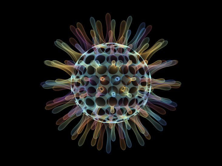
COVID, like all other viruses, attacks primarily the Mitochondria and DNA of cells see figure [adapted from [i]].  It attacks the DNA to help them replicate.  In this way, the virus is able to produce the proteins that it needs to live.  It cannot do this on its own.  This is one reason why viruses are very species-specific[1].  As parasites, they attack the mitochondria to obtain the energy they need to live by siphoning off a significant portion of the energy molecules (called ATP – adenosine tri-phosphate) that we also need to be active or even live.  The virus, by infecting us, leaves less energy for us the host.

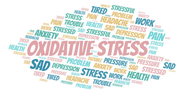
Nerve cells and heart muscle cells contain, by far, the most mitochondria in the human body.  This attack on the mitochondria results in what is known as oxidative stress.  Oxidative stress[2] causes fatigue, exercise intolerance, and many of the other mental, cognitive, and physical (including pain) symptoms reported with PACS.  In most cases, it is like having a full fuel tank in your car with a clogged fuel filter.  You seem fine at rest (while idling), but as soon as you step on the gas to go (and be active), nothing happens because the necessary, additional gas (energy molecules) does (do) not reach the engine (the brain and nervous system and the heart).  In this case, the extra energy molecules are actually not available because they have been stolen by the virus.  All parasites (viruses, bacteria, molds, mildews, etc.) affect the mitochondria and energy supply this way.  This is why you are fatigued and have no energy when you are sickened by these agents.  Add to this the fact that COVID also compromises the lungs, reducing the host’s oxygen intake, causing less fuel to generate energy molecules, and you have the SARS effect.

One cause of Oxidative Stress is indeed cytokine dysregulation.  The “Cytokine Storm,” referenced as a “Viral Tornado” in the 20/20 interview of Mount Sinai Hospital in November 2020, is due to oxidants, including reactive oxygen species (ROS).[3]  Cytokine storm and oxidative stress are the main players of Acute Respiratory Distress Syndrome development during respiratory virus infections.[4]  Mechanisms of cytokine production (cytokine storm) and epithelial barrier disruption by respiratory virus infection leads to the enhanced ROS production.  

Cytokine Storm and Oxidative Stress in COVID-19 patients result in a vicious cycle[5], causing Hypercytokinemia, known as cytokine storm[6].  Given this, simple therapies to prevent oxidation and excessive cytokine release (Hypercytokinemia) are prevalent and readily available.  In fact, some are already being used, including Vitamins C & D and Zinc, tacitly confirming the Oxidative Stress hypothesis.

If the politically motivated powers that be would simply admit this commonly and well-known fact, Oxidative Stress is readily treated with antioxidants.  Again, that is, in large part, the reason why Vitamin C & D and Zinc are a significant part of COVID therapy.  The problem for the politicals is that these therapeutic agents do not make anyone any money.  They have to fear the public into believing that special medications and vaccines are required.  This is why commonly used and effective medicines that have worked here in the US and elsewhere around the world were pulled from doctors, because, again, they did not make any of the political powers any money.

The effect on nerve cells, especially Parasympathetic and Sympathetic (P&S) nerve cells, is what eventually leads to post-COVID syndrome[7].  These authors wrote an Op/Ed back in March of 2020 stating that a post-COVID syndrome will present.  We still cannot get that manuscript published (over 10 journals have refused to print it – that is why we have had to create our own journal and print it here in our own journal).

Oxidative Stress and its effect on the P&S nervous systems explain the first issue of PACS, a potential delay in the symptoms.  Initially, this was a problem of doctors, and may still be a problem based on a recent Op/Ed[8], writing about “echoes of chronic fatigue in the effort to blame the coronavirus for a host of questionable symptoms.”  This delay seems to separate COVID from PACS.  Add to this the general lack of knowledge about the P&S nervous systems, and this sort of ignorance gains credibility.  Especially when the patients seem normal at rest, for when most tests are administered, the patient is either sitting or lying down.  However, the P&S nervous systems are rarely resting.  In fact, when you are resting is, arguably, when the P&S nervous systems are most active.

The P&S nervous systems control or coordinate all organs and organ systems within the body (including themselves).  The job of the P&S nervous systems, collectively, is to maintain normal organ function, even though they themselves may not be functioning normally.  Until the P&S system fails, the organs remain functioning normally, unless they are attacked directly.  Once the organs fail, then symptoms present.  It is the unhealthy or improperly controlled organs that generate symptoms.  The delay in symptoms presenting is based on how long it takes the P&S systems to fatigue or become significantly un-balanced (fail), permitting the organs to then function abnormally. resulting in symptoms.

It is like the heating and cooling systems of your house.  Assume you have no knowledge of, nor are able to see, hear, or access the cooling and heating systems of your house, and that they are individual systems working together to keep you comfortable.  Assume all you have to assess the function of those systems is the thermostat in the house.  Say you set the temperature on the thermostat to 70°F (like 70 beats per minute for heart rate).  Assume it is a hot, humid summer day in the south somewhere.  Assume that the cooling system is well.  Now assume that a $5.00 heater switch fails and the heater turns on to full capacity.  What happens to the temperature of the house as measured by the thermostat?  …  NOTHING!  …  The cooling system amps up to compensate not only for the ambient heat but also for the heat from the heating system.  Now both systems are operating at full capacity or more, and the thermostat temperature still reads 70°F, and everyone is still happy.

How long will this situation last until there is a catastrophic failure in one or both of the air conditioning systems?  Only after a catastrophic failure does the temperature change.  Then it is too late, and the repair is $5,000.00 or more.  The change in temperature, indicating a failure, is too late!  The $5.00 repair is simpler, and easier, but you have to be measuring both the cooling and heating systems, not just the net result of their combined activities.

The autonomic nervous system in the human body is very similar in this regard.  We are still surprised by heart attacks and strokes and other organ failures because we only measure the autonomic nervous system (the net result of the P&S nervous systems) and not the individual P&S nervous systems themselves.  This is made more poignant by the fact that it is the purpose of the P&S nervous systems to work together to maintain normal organ function, even when they themselves are dysfunctional.  Current, common-place healthcare measurements are made more effective and efficient with the additional information of P&S Monitoring.

The second issue is that the parasitic effect of COVID is on all mitochondria within all organs.  This compounds the effect of an unbalanced P&S nervous system and the inefficient control from them, which also affects all organs. 

Again, unfortunately, relatively little is known about the P&S nervous systems because, until rather recently, an efficient and simple method of measuring it was not widely accepted, and the more widely known measures of the Autonomic nervous system as a whole were not able to specify the underlying dysfunctions sufficiently so as to develop effective treatments or to demonstrate that treatment was even possible. 

This leaves doctors with the current plan of simply “characterizing the disease.”  In other words, simply listing all of the symptoms and simply treating all of the symptoms individually.  This of course leads to many prescription medications, whose multiple interactions are unknown, perhaps causing more symptoms than they (collectively) are supposed to relieve. 

In fact, this compounding effect of unbalanced P&S control of weakened or dysfunctional organs multiplies the number of symptoms and adds more disorders, some of which appear to be mental illnesses as well.  Note, most of these mental illnesses are also explained by unbalanced P&S control causing reduced blood flow to the heart and brain, resulting in brain fog, cognitive and memory difficulties, depression, anxiety, headache and migraine, muscle and joint pain, and sleep difficulties, as well as the fatigue, lightheadedness, and malaise commonly associated with PACS.

The lack of understanding of the P&S nervous systems leads to a third general problem or issue.  Without the additional information from P&S testing, medicine has no other choice but to simply treat symptoms (which plays into the hands of the pharmaceutical and marijuana industries, enabling them to sell more product).  Yes, understanding the P&S nervous systems does explain all symptoms of COVID and PACS,   including Diarrhea.[9],[10] 

Only the P&S nervous systems connect all symptoms and organ systems involved in the COVID and PACS conditions.  In the example of Diarrhea, yes, the virus is in the stool, but then all viruses end up in the stool, so what is special about COVID? COVID strongly affects the immune system, which causes Parasympathetic Excess, which overdrives the motility of the GI system and leads to Diarrhea.  Let’s not trample the issue out of ignorance.  Let’s get to the underlying cause, do our best to minimize mortality risk and target the cause to minimize morbidity risk.  Then, treat the end-organs that were themselves damaged and remain dysfunctional.

Depression (and Anxiety) are also more likely to be a result of dysautonomia.  If the brain receives low levels of blood flow due to at least three types of dysautonomia, some of which may present simultaneously, the brain will be “asleep” while the patient is upright, sitting or standing, all day.  A brain “asleep” is too often misdiagnosed as a brain “depressed.” 

Then, if the blood flow drops any lower (due to a large meal or emotional event, etc.), the brain will issue an “Adrenaline Storm,” which is its way to call for more blood.  The Adrenaline Storm may then cycle Anxiety.  Treating this Depression/Anxiety condition with high dose antidepressants, such as Fluvoxamine (an OCD drug)[11]m ay, in some people, help to increase blood flow to the brain through other P&S interactions, but this is a very inefficient therapy and may be a self-fulfilling prophecy, forcing the patient to think that they have a mental health issue.  Besides, the data are scant, and the conditions are not well controlled to rule out isolation, masks, and social distancing, which are contributing enough to Depression.  We do not need any other causes.

Unfortunately, in many cases, simply increasing the numbers of medications and their dosages, in the hopes of relieving the symptoms faster, actually leads to more complications.  Western pharmaceutical theory is based on the understanding of what one chemical (as a medication) in isolation will do to the human body.  The interactions between more than two or three chemicals (medications) are not well known.  This is the main reason why the very ill or elderly who are on 15 to 17 medications a week are chemically relegated to very poor qualities of life, sitting as lumps in their wheelchairs with no energy of motivation to be productive.

It is like the old nursery rhyme of the old lady that swallowed a fly, then a spider to get the fly, then a cat to get the spider, then a dog, etc.  By the time you get to swallowing the elephant and the doctor is considering prescribing you a blue whale, it may be prudent to consider getting the fly.

The “fly” in the case of COVID is Oxidative Stress, which may be “gotten” with the super antioxidants, r-Alpha-Lipoic Acid (rALA) and Co-Enzyme Q10 (CoQ10).  rALA is selective for nerves and heals the mitochondria, DNA, and other cellular processes that are affected by the virus, and CoQ10 is selective for heart muscle cells effecting the same repairs in those cells.  Furthermore, rALA & CoQ10 are the most powerful antioxidants the body produces (second only to the most powerful antioxidant of all, which is exercise), and they make themselves even more powerful by recycling other antioxidants, such as Vitamin C & D, and Zinc.

So, why are we grasping at straws?  Again, in the hysteria, we are overlooking the simple solutions.  Unless of course it is because the simple solutions do not make the instigators of all of this enough money.  Now there is a KF-94 mask that is 94% effective, as opposed to the KF-95 mask that is 95% efficient in filtering out airborne particulates.  The article[12] states that the bottom line is this: “It’s not always just about filtration efficiency.”  The article continues to state that these (surgical-grade) masks, such as the KF-94, are for use in a hospital because they’re designed for medical settings.  In the real world, it may be they are hard to wear through the course of the whole day.  The best mask is the one you can wear all the time.

So, is it about filtration or comfort?  Viruses are very, very small!  Are we trying to prevent the spread of the virus or not?  Anything but a surgical-grade mask is useless in preventing the spread of the virus because the virus goes through the mask as if it were not there, and even with a surgical-grade mask, by the time a man has “5 o’clock shadow,” his hair has pushed the mask off his face far enough for the virus to escape un-impeded; not to mention men with beards.  Similarly for some women with make-up, the microscopic peaks and valleys may be large enough for the virus to fit through and escape.

Another article[13] is entitled “International team of scientists identifies new treatment for COVID-19 that appears to be far more effective than drugs in use now.”  However, it has only been tested on cells in the lab.  The article concludes that “Even though ‘a drug is effective in cells in the laboratory, we don’t know what effect it will have on cells in the human body.’”  So, how is this a new treatment yet, let alone better than current drugs?  Again, science needs to be left alone to do their work and not give false hopes.  Similarly, another article states that a “new Israeli drug cured 29 of 30 moderate/serious COVID cases in days in hospital.”[14]  Great, 29/30!  But how were the patients picked?  What were their histories?  There are still a million questions to be answered.  There is a reason why it takes years and 10,000 patients to approve a new drug.  What happened to the one that it did not cure and why?  Did that patient die?  A death rate of 1/30, or 3.33% is an awfully high death rate for a new drug.  Killing the patient is not an acceptable side effect.

However, just relieving Oxidative Stress does not always repair the damage done to the P&S nervous systems.  Further therapy may be required to rebalance the P&S systems to return health and wellness.  P&S imbalance must be measured and monitored, because everyone is different, and the individual’s P&S systems at that time must be known in order to plan therapy.  Some consider the P&S nervous systems as your physiologic fingerprint.  They include and remember your entire medical and life history.

There are at least four P&S dysfunctions (dysautonomias) that may precipitate the symptoms of post-COVID syndrome, and more than one is quite possible, and their specific interactions are based on the individual patient’s own clinical and personal histories.  All four dysautonomias, inappropriate P- or S-actions, individually explain the majority of PACS symptoms.  Combinations of the four, which are more likely, in our experience, explain the range in severity of PACS.  For more information, visit www.physiops.com.

Treating the P&S nervous systems, however, is rarely a fast process, nor does more medication hasten the process; in fact, quite the opposite.  Much like a pendulum, hitting it hard knocks the pendulum off its hinge and you have many more problems.  Treatment requires small, gentle, easy nudges over time to correct “the pendulum.”  So, it is with the P&S nervous systems.  Further, it is not like the cold where you get ten days of therapy and are healthy again.  It may take up to three months to effect a change in the P&S nervous systems.  It is more like breaking a bad habit and establishing a new, hopefully good habit.  Even still, there are few if any changes in symptoms because the organs have yet to change.  Changing the organs may take another three months depending on the severity of the insult.  These time frames are also lengthened by age and severity of the insult, in this case, COVID.  Patients must be diligent and patient during these six months or the condition will worsen, and it may take years to recover.

The average patient, by the time they visit a P&S clinic, have seen dozens of doctors over decades of time.  The problem is that the other doctors only assess them when they are at rest, sitting or lying down.  Rarely do they assess them while active.  As the typical test results show, they are normal at rest – which is why many are not believed.  They had better be normal at rest – they have had numerous doctors working really hard to make them normal … at rest; as attested to by P&S monitoring results.  Again, a problem is that this portion of the nervous system never rests.  Therefore, the P&S must be tested while active as well.  This is the basis of P&S testing and always shows the reason(s) for a patient’s symptoms, as well as morbidity and mortality risks (respectively, they are the risks of additional symptoms, which is morbidity risk, and the risk of a major adverse event, including heart attack, stroke, and death, which is mortality risk).

The fourth issue, again based on the lack of understand and data on the P&S nervous systems, is that physicians have been left with few therapy options that are specific for the P&S nervous system.  Unfortunately, there are, in fact, only two medications approved for P&S dysfunction, and both are approved for the same, singular dysfunction.  Fortunately, it is a common P&S dysfunction that is common to post-Oxidative Stress conditions:  Orthostatic Dysfunction, typically Postural Orthostatic Tachycardia Syndrome (POTS) or Orthostatic Hypotension.  All other therapies are applied off-label.  This causes other problems, including the fact that in western medicine, titrating patients to high levels of medication is common.  This desensitizes patients to the low dosages needed to treat the P&S systems and not cause more symptoms.  This often forces physicians to use supplements and lifestyle modifications, which are often less reliable or effective; especially if Oxidative Stress is not considered.

Given all of this, you begin to see why medicine is more focused on simply treating the symptoms of post-COVID syndrome or PACS, and then trying to convince you that this is your new normal and you must embrace it.  Even before COVID, we have been railing against that philosophy, because as tens to hundreds of thousands of P&S (not autonomic, but P&S) patients across the country will attest, it does not have to be so – you may have a decent quality of life and productivity restored.


[1] As an example, all human HIV studies were performed on simian (monkey) HIV viruses so enable safety in the lab and not infect the humans doing the research.

[2] PubMed, a very large collection of highly reputable medical journals, alone lists 273 references associating coronavirus, specifically, with oxidative stress.  This does not include the thousands of references associating other viruses with oxidative stress.

[3] Ye S, Lowther S, Stambas J. Inhibition of reactive oxygen species production ameliorates inflammation induced by influenza A viruses via upregulation of SOCS1 and SOCS3. J Virol. 2015;89(5):2672-2683. doi:10.1128/JVI.03529-14

[4] Meftahi GH, Bahari Z, Jangravi Z, Iman M.  A vicious circle between oxidative stress and cytokine storm in acute respiratory distress syndrome pathogenesis at COVID-19 infection .  Ukr.Biochem.J. 2021; Volume 93, Issue 1, Jan-Feb, pp. 18-29.  doi:https://doi.org/10.15407/ubj93.01.018

[5] Khomich OA, Kochetkov SN, Bartosch B, Ivanov AV. Redox Biology of Respiratory Viral Infections. Viruses. 2018; 10(8):392. https://doi.org/10.3390/v10080392

[6] Suzuki K. Cytokine Response to Exercise and Its Modulation. Antioxidants. 2018; 7(1):17. https://doi.org/10.3390/antiox7010017

[7] PubMed alone lists over 1200 references associating oxidative stress with P&S dysfunction or imbalance.

[8] The Dubious Origins of Long Covid, by Jeremy Devine  March 22, 2021 6:36 pm ET, Wall Street Journal

[9] Coronavirus symptoms: This is the likely order in which COVID-19 symptoms appear after you get infected, by Anushree Gupta  Updated Feb 01, 2021 | 16:14 IST, https://www.timesnownews.com/health/article/coronavirus-symptoms-this-is-the-likely-order-in-which-covid-19-symptoms-appear-after-you-get-infected/714563

[10] COVID-19 and Diarrhea, https://www.news-medical.net/health/COVID-19-and-diarrhea.aspx

[11] What Is Fluvoxamine? OCD Drug Could Be Used to Treat COVID, by Jason Murdock On 3/8/21 AT 9:49 am EST

[12] “Coronavirus FAQ: Why Am I Suddenly Hearing So Much About KF94 Masks?” by Pranav Baskar  January 22, 20216:12 pm ET, https://www.npr.org/sections/goatsandsoda/2021/01/22/959683338/coronavirus-faq-why-am-i-suddenly-hearing-so-much-about-kf94-masks

[13] International team of scientists identifies new treatment for COVID-19 that appears to be far more effective than drugs in use now, by Mark Johnson  Milwaukee Journal Sentinel https://www.jsonline.com/story/news/2021/01/25/international-team-finds-new-more-effective-drug-treat-covid-19/6673529002/

[14] New Israeli drug cured 29 of 30 moderate/serious COVID cases in days — hospital, by Toi Staff 5 February 2021, 3:29 pm, https://www.timesofisrael.com/new-israeli-drug-cured-moderate-to-serious-covid-cases-within-days-hospital/

[i] Rasa S, Nora-Krukle Z, Henning N, Eliassen E, Shikova E, Harrer T, Scheibenbogen C, Murovska M, Prusty BK; European Network on ME/CFS (EUROMENE). Chronic viral infections in myalgic encephalomyelitis/chronic fatigue syndrome (ME/CFS). J Transl Med. 2018 Oct 1;16(1):268. doi: 10.1186/s12967-018-1644-y. PMID: 30285773; PMCID: PMC6167797.

Read More

How hypermobility Ehlers-Danlos Syndrome affects Parasympathetic and Sympathetic (P&S) System?

Click here to download this post 

DePace NL, Acosta CR, DePace Jr. NL, Kaczmarski K, Goldis M, Colombo J.

What is ehlers danlos hypermobility syndrome?

Hypermobility/Ehlers-Danlos Syndrome (hEDS) defines a spectrum of connective tissue disorders that are caused by defects in the genetic information that is used in humans to produce collagen. 

In both, the collagen is long and flexible, rather than short and stiff.  This results in loose and “leaky” connective tissue.  hEDS may be inherited, usually an autosomal dominant trait, however acquired cases occur frequently. 


What are the characteristics of Hypermobility Ehlers-Danlos Syndrome

To date there is no known cure for hEDS (Hypermobility Ehlers-Danlos Syndrome). However, there are a few characteristics of Hypermobility Ehlers-Danlos Syndrome that are well known (in no particular order): 

1) hEDS affects females significantly more than males;

2) In the young, the additional flexibility seems advantageous due to the lack of significant symptoms;

3) Generally, around the end of development (during later teens or early 20s) symptoms begin to present, and generally active and vivacious teenagers become sickly for no apparent reason with poor and, frequently, debilitating qualities of life.

Another primary characteristic of hEDS patients is that they demonstrate some degree of autonomic dysfunction (a.k.a., dysautonomia).  This may explain the last two characteristics listed above. 

When Dysautonomia Symptoms Appear?

During development, except for a couple of years around ages 8 and 15 when development slows down, the autonomic nervous system (ANS) is very active in development; therefore, dysautonomia symptoms are masked and the symptoms that appear are attributed to current factors with largely unknown histories. 

Once development ends, in the late teens or early twenties, dysautonomia symptoms are unmasked and the effects of a persistently overactive ANS presents.  In general, dysautonomia is the effects of an imbalance between the two autonomic branches:  the Parasympathetic and Sympathetic (P&S), nervous systems. 

What does Research Say About Parasympathetic and Sympathetic (P&S) Activity in Hypermobility Ehlers-Danlos Syndrome Patients?

Here we introduce a clinical cohort and some general characteristics of P&S function in 243 patients, predominantly female.

Younger hEDS patients’ P&S activity is high-normal possibly due to their heightened immune state and their body’s attempt to heal the “leaky” connective tissue.  The initial high-normal S-levels, higher than the P-levels (the opposite is typical), may be why there is persistent inflammation starting in the earlier years, a characteristic of hEDS patients. 

Typically, given that P-activity is more involved in development and pregnancy, P-activity is higher during these years, as in the normal subjects through the 20s and 30s.  Allergies, Mast Cell activation, Arthritis, Small Fiber disorder, etc. all involved S-activity. 

Histaminergic and inflammatory responses are Sympathetic functions.  As the Sympathetics are the reactionary branch, S-activity is normally short-lived. 

Persistent or inflated S-activity, therefore, leads to histaminergic and inflammatory disorders.  In hEDS cases, S-activity is typically inflated by the elevated P-activity and the additional S-activity drives the additional inflammation. 

S-activity is also involved in the pain response.  Amplified S-activity, due to abnormal, excessive P-activity[1], also amplifies the pain response, especially in “Fibromyalgia-like” pain syndromes. 

Since the majority of hEDS patients are female (91.8% of this total population), P-activity (and therefore S-activity) remains higher during childbearing and may be the reason for the persistence of the elevated P&S activity into the middle-age years.  Subsequently, the hEDS patients’ resting P&S activity normalizes, as compared with that of the Normal subjects.  However, the hEDS patients’ resting S-activity remains high compared with resting P-activity.

hEDS is believed to not be life-threatening, except for one form of EDS (the vascular form).  However, based on these sample populations, hEDS may perhaps reduce length of life an average of 10 years; given that they cross the CAN threshold up to 10 years earlier than Normals. 

Again, P-activity is protective.  The more reduced P-activity in the hEDS patients may be the cause of the reduced life span.  It may indicate an immune system that has fatigued earlier or it may be responsible for earlier onset MACE-risk (heat attack, stroke, heart failure, etc.). 

The higher mortality risk is reflected in the CAN with SB > 2.5 condition in the hEDS patients starting around age 60, on average.  CAN with SB > 2.5 is therefore an indicator that therapy should be more aggressive about establishing and maintaining low-normal SB:  0.4 < SB < 1.0; e.g., increasing dosages of Sympatholytics or reducing stress, including Psychosocial stress.  Overall, the P&S data are a better match to the natural history and progression of hEDS.

Many patients present with prior diagnoses of depression and anxiety or psychiatric illness attributed to them, but they know they have something real and abnormal that is not purely psychiatric.  The patients hurt all over and have diffuse pain, which keeps them from functioning properly.  They are often diagnosed as “Fibromyalgia” or “Chronic Pain Syndrome.” 

Many cannot perform any gainful employment.  Certainly, they become anxious and depressed because of their non-functional status.  Dysautonomia features, such as exercise intolerance, orthostatic intolerance (where one cannot stand up without getting brain fog or dizzy), and chronic or persistent fatigue are almost universally present in these patients.  There is a high percentage of females with this problem, but we do also see males in addition, since it is believed that if a person has this disorder, they can transmit it genetically to one or two of their children (autosomal dominant transmission).

It is well known that the P&S is, generally, very active during development.  This level of activity, masking any effects of excessive P&S activity, may explain why symptoms do not present until after development.  It has been postulated that “leaky” connective tissue permitting foreign items to leak-in causes a persistently, heightened immune response. 

This in-turn leads to a persistent state of Parasympathetic Excess (PE), mostly while active as well as for periods of time when younger at rest, as measured as high-normal SB (2.0 < SB < 3.0).  The persistent and prolonged stress on the more exposed and longer Parasympathetics (Vagal) nerves, including Oxidative Stress, tends to cause them to weaken first and fastest. 

The relative PE also forces a relative Sympathetic Excess (SE) which not only multiplies symptoms, but amplifies symptoms, such as Sympathetically-mediated pain and inflammation. 

Unfortunately, there is no cure for P&S imbalance in these patients.  Typically, with many other diseases and disorders, once P&S balance is established, then the nervous system “learns” this new condition and maintains it until some other clinical event occurs.  Unfortunately, in hEDS, the next clinical event, per se (such as the next infection), is only moments away once it leaks into the body. 

Therefore, there is also no real cure for P&S imbalance due to hEDS, only the ability to treat it to maintain P&S balance as much as possible as the systems continues to degrade more rapidly than normal.  Fortunately, once the protocol for the individual patient is determined, it may be implemented immediately should a significant clinical event occur, including pregnancy where patients may need to suspend treatment during that time.

Fatigue, exercise intolerance, shortness of breath, palpitations, and chest pains with which many patients with hEDS present, are often the result of P&S dysfunction. 

This goes hand in hand with the hEDS.  Even the amplified and generalized pain and inflammatory responses (not only in the joints), anxiety, brain-fog, memory and cognitive difficulties, sleep difficulties, GI motility issues, may be secondary to P&S dysfunction caused by hEDS. 

Many patients develop Small Fiber Disease which is an inflammation or dysfunction of unmyelinated small, type C nerve fibers which carry autonomic and sensory, including pain, signals. 

Also, hEDs is associated with Mast Cell hyperactivity which manifests as episodic histaminergic over-production.  Mast Cell may be associated with Celiac disease and food allergies or sensitivities.  Leaky Gut Syndrome may be involved either as a result of histaminergic excess or leaky connective tissue. 

Histaminergic over-production may be associated with persistent or excessive Sympathetic activity secondary to PE.  This may be tested for objectively and serially, with diagnostic test modalities that provide quantitative information.  This, in-turn permits more individualized titration of therapy given the then current state of the patient’s nervous system.

One reason for the lack of understanding and recognition of the P&S dysfunctions underlying hEDS is the fact that virtually all of the data collected from patients are collected while the patient is at rest. 

The ANS, specifically the P&S nervous systems are never at rest. (In fact, it may be argued that they are most active when you are sleeping, resting.)  As a result, the common thread behind the constellation of symptoms associated with hEDS is lost. 

It is well known that the Sympathetic nervous system is the reactionary branch of the ANS and is not supposed to be chronically active.  It is also well known that the Parasympathetic nervous system is the ANS branch that establishes the metabolic threshold around which the Sympathetic branch reacts and then works to quiet the Sympathetic branch.  Under normal resting conditions as one branch is activated the other becomes less active.

This is not the case in most abnormal conditions and is not the case under abnormal dynamic conditions.  There are two significant, dynamic P&S abnormalities (P&S dysfunctions that present when not at rest) that are, apparently, caused by hEDS and serve to exacerbate the symptoms of hEDS. 

One is known as Sympathetic Withdrawal (SW), which is an abnormal alpha-Sympathetic response to head-up postural change (sitting or standing) which leads to poor cardiac and cerebral perfusion which lead to fatigue, exercise intolerance, shortness of breath, palpitations, and chest pains, and anxiety, brain-fog, memory and cognitive difficulties, and sleep difficulties; respectively.  The other is known as Parasympathetic Excess (PE).which is an abnormal Parasympathetic response to a stress (a beta-Sympathetic) response. 

PE not only may exacerbate symptoms caused by SW, but it also amplifies the Sympathetic disorders, including pain and inflammatory responses (including in the joints).  It may also cause, or be the cause of, Mast Cell hyperactivity leading to unexplained rashes and Mast Cell Activation Syndrome (MCAS), and Small Fiber disorder.

PE may be a primary disorder caused by hEDS.  It is well known that the Parasympathetics control and coordinate immune responses, including providing the “memory” for the immune system.  Since hEDS enables foreign substances to “leak-in” to the body all of the time, the immune system is always on constant, heightened “alert” and thereby forces the Parasympathetics to remain overactive. 

Dynamically, PE forces the Sympathetic response to also be excessive (Sympathetic Excess or SE), secondarily.  Unfortunately, since most clinical office measurements are Sympathetically-based (HR, BP, etc.), only the SE is recognized (high HR and high BP), and therefore treated. 

However, this results in more PE because the complimentary Sympathetic activity is reduced enabling the increase in PE.  This often leads to unresponsive or labile patients which are often misinterpreted.  In these patients, when PE is recognized as a primary autonomic dysfunction, and treated as such, the secondary SE is often relieved organically, in time, and then the Sympathetically-based symptoms are often relieved organically, in time, assuming no end-organ effects.

A final thought for now.  Since the human body will assimilate foreign, ingested, collagen (i.e., from bones, shellfish shells and other animal connective tissue) perhaps this is a basis for some relief of hEDS.  The (normal) animal collagen may help to “plug the leaks” caused by the abnormal native collagen.  Many patients empirically find relief of symptoms with intake of collagen products.

As mentioned before, there is no genetic testing or lab testing that is diagnostic of hEDS.  That is not to say that we will not in the future hone down on a specific gene loci or other biomarkers that may be supportive of hypermobile Ehlers-Danlos Syndrome.  However, to date, there are none. 

Franklin Cardiovascular uses a scoring system developed by the EDS Society.  For now, it is possible to re-establish P&S balance and thereby help to restore an improved quality of life that permits a productive lifestyle, with less pain and better sleep.  This is just the beginning of a research effort that must include many more patients from many more sources as hEDS awareness continues to grow.

Read More

Do I Have Mast Cell Activation Syndrome (MCAS)?

Click here to download this post 

Mast Cell Activation Syndrome (MCAS) is a disorder where components of the blood stream, namely mast cells, secrete various substances which can be involved in an allergic reaction or inflammatory reactions.  However, before discussing MCAS, we need to understand what the mast cell is and where it comes from.

What is Mast Cell?

Mast cells come from more undifferentiated-type cells in the bone marrow.  They usually mature in various tissues.  Mast cells are important reactionary cells in allergic reactions and in inflammatory reactions.

They secrete substances, such as Histamine, Prostaglandins, Leukotrienes, various enzymes which can break up other substances known as Proteolytic  enzymes and Cytokines, such Interleukins 6, 18 and 13.  Also, Tumor Necrosis Factor (TNF) and Vascular Endothelial Growth Factor (VEGF) may be secreted.  These substances may cause inflammation and also activate the immune system in various circumstances. 

Normally, mast cells do not spontaneously secrete these substances but in disorders such as MCAS they can.  Common triggers for mast cells to secrete these substances include IgE and antigens during an allergic reaction, anaphylatoxins, Cytokines, hormones, and substances such as Substance P (SP).  In fact, SP may be the main trigger in many skin disorders such Contact Dermatitis, a disorder in which mast cells are activated and secrete many of the substances named above.  In addition to Contact Dermatitis, Mast cells are very importantly involved in many other skin abnormalities, immunological responses, gastrointestinal responses, and may, interact and affect virtually every organ in the body.

What is Mast Cell Activation Syndrome

MCAS is a chronic condition involving multiple organs in which normal mast cell activation leads to the inflammation and allergic symptoms that may occur episodically in patients.  Gastrointestinal symptoms are common including Irritable Bowel Syndrome

Recently, the term mast cell activation syndrome disease (MCAD) has been defined.  This is the major heading for MCAD with two main categories. 

One is known as Systemic Mastocytosis (SM) and MCAS. 

Both of these disorders may have similar symptoms and systemic manifestations.  Usually with SM and its subclass, Mast Cell Leukemia (which is very rare), there is a genetic or clonal abnormality and there is usually an abundance of mast cells produced or a higher quantity exists; whereas, in MCAS, the number of mast cells are not increased, they are only hyperactive.   It is not known if MCAS can be transferred over time into the rare neoplastic or malignant states of SM and Mast Cell Leukemia.

What Causes Mast Cell Activation Syndrome

The triggers of MCAD include stress, food, alcohol, and various medications including possibly aspirin, infections, air pollution, heat, mold, chemicals, and changes in our intestinal microbiome.  The latter may be affected by antibiotics or stress.

What is the definition of MCAD?  Over the last ten years, much has been devoted towards establishing a clear definition for this disorder. 

Criteria have been proposed, and three criteria are specifically agreed upon.  It is important to satisfy all three criteria before concluding that the given patients’ symptoms are due to mast cell activation. 

It should be recognized that idiopathic anaphylaxis is a specific entity within the MCAS.  A patient may, however, experience urticaria or hives or gastrointestinal symptoms after exposure to a possible trigger allergen.

Mast Cell Activation Syndrome Symptoms

While many of the symptoms of MCAS (see below) are nonspecific in nature, again, there are specific criteria that must be fulfilled before one can diagnosis a patient as having MCAS.  There have been many criteria, but the ones most commonly used require symptoms consistent with chronic recurrent mast cell release.  These include:

  1. Recurrent abdominal pain, diarrhea, flushing, itching, nasal congestion, coughing, chest tightness, wheezing, lightheadedness, or a combination of some of these.
  2. Laboratory evidence of a mast cell mediator (elevated Serum Tryptase) whether at baseline or with provocation or during an attack, N-methylhistamine, Prostaglandin D2, or 11-Beta-prostaglandin F2 alpha, Leukotriene E4 and other mediators as determined by various laboratory measurement that pertain to mast cells.
  3. Improvement in symptoms with the use of medications that block or-treat elevations in these mediators, specifically Histamine blockers and other mast cell stabilizers.

Sources of MCAS Symptoms

Symptoms of MCAS can derive from any organ system and one usually needs two organ systems or comorbidities of at least two organ systems to fulfill criteria #1 above.

In regard to constitutional symptoms, fatigue and weakness, heat and cold sensitivities and sleep deprivation are commonly identified.

Dry eyes, red itchy and red burning, runny nose, and inflammation ulcers of the mouth may be seen in the head and neck organ system.

In regard to the chest and heart, chest discomfort, rapid heartbeats, redness, flushing of the skin, sudden dizziness, hot flashes, and blood pressure surges may be seen.  Also, syncope and presyncope.

In regard to the pulmonary system, dry cough that occurs repeatedly, shortness of breath, difficulty taking a deep breath, and episodic asthma and wheezing-like complaints can be present.

For the gastrointestinal system, abdominal symptoms are common to include pain, crampy or spastic discomfort oftentimes associated with diarrhea, abdominal bloating and distention, and symptoms of irritable bowel syndrome and diarrhea is also noted.  Swallowing difficulties and throat tightness are also noted.

In regard to the urinary tract and pelvis, bladder and pelvic pain as applies to both men and women may be present.  There may be painful, frequent and urgent urination or pain during sex.  The disorder of Interstitial Cystitis has been described where it is believed mast cells are very operative in its presentation and where an individual has significant urinary tract symptoms and discomfort, but does not have a documented urinary tract infection.

Neurological symptoms may occur with headaches, brain fog and neuropathic leg or arm pain.

The skin is one of the most affected organ systems by mast cells.  Hives, itching, swelling of the lips, cheeks, eyelids, reddish-brown spots under the skin and occasional hemangiomas are noted.  One may see reddish or pale complexion, itchiness with a burning feeling, and Dermatographism is common.

In regard to the hematologic system, one can see bruising and unusual nose bleeds.

In regard to the bones, patients can demonstrate bone pain.

Also, immune system involvement can be noted.  There have been immunological disorders, such as Common Variable Immunodeficiency Syndromes associated with MCAS.  One needs to determine if they get head colds or upper respiratory infections frequently and if they turn into bacterial infections, such as bronchitis and sinus infection which are common, and do these infections come on with attacks episodically that are related to mast cell activation.

How is Mast Cell Activation Syndrome Diagnosed

Various physicians will order different tests to determine if there is an increase in mast cell mediators. 

Various Mast Cell Mediators

Oftentimes all of these tests can come back negative for MCAS, but during attacks if these mediators, specifically Serum Tryptase, are tested during the first 1-4 hours, we can see a rise above baseline and can confirm objective data to support their diagnosis. 

Serum Tryptase

As mentioned, Serum Tryptase is an important mediator, and during an attack one likes to see at least a two-fold plus 20% increase in this value to consider that significant.  At times, Tryptase will be elevated at rest, and if it is above six (6.0), one may have to look towards a genetic enzyme abnormality. 

Histamine

Histamine can be measured in the plasma and its metabolite N-methylhistamine can be measured in the urine, and plasma histamine in the blood.  We often like to see this number more than 10 times the upper limit of normal, but any elevation is important. 

Prostaglandin D2

Prostaglandin D2 in the plasma is also measured as Heparin or Factor 8.  Chromogranin A, which is nonspecific and can be seen in neuroendocrine tumors and other gastrointestinal disorders or can be elevated in renal failure. 

If increased, it is very suspicious for MCAS in patients who do not have the former disorders.  The Leukotriene E4 in urine is also an important mediator to test for. 

Another important mediator to test for in the urine is PG-D2 or 11β PG F2α.  In addition, many times a biopsy is taken of the skin or the GI tract during endoscopy or colonoscopy. 

Other Tests for MCAS

If focal or disseminated infiltrates or morphologically inconspicuous mast cells are seen, or a mast cell collection, or a morphology of spindle shaped mast cells or if they are specially stained for CD25-positive mast cells, this gives significant strength to the diagnosis of MCAS.

One has to exclude other disorders which may mimic MCAS to make sure the symptoms are not due to Diabetes, Porphyria, Thyroid diseases, Amyloidosis, Hepatitis, Gallbladder disease, infectious Enteritis, Carcinoid tumors, Pheochromocytoma, (a tumor of the adrenal gland which can elevate blood pressure), pancreatic endocrine tumors, Eosinophilic Syndrome abnormalities, hereditary Angioedema, Vasculitis and rarely, intestinal Lymphomas.

Mast Cell Activation Syndrome Treatment

Treatment of MCAS or suspected MCAS is important because a response fulfils one of the criteria above.  Usually we begin with H1-antihistamines, such as Cetirizine (Zyrtec*), Ketotifen (Zaditor), or Fexofenadine (Allegra) or Loratadine (Claritin). 

H2-histamines, such as Famotidine (Pepcid*) or Ranitidine (Zantac) are added on.  This is usually first-line treatment using both an H1 and an H2 agent.  If the response is not complete, we often go to Antileukotrienes, such as Montelukast (Singulair) or Zileuton (Zyflo). 

Some people use natural products, such as Curcumin or St. John’s wart.  If not contraindicated, or not determined to be a triggering agent, a nonsteroidal anti-inflammatory (NSAID) agent and aspirin can be helpful in reducing inflammation in some of the patients. 

Oftentimes, we will tailor the therapy if a certain mediator is tested for and is elevated in the urine or blood.  For example, Prostaglandin elevation may influence us to use nonsteroidals or aspirin earlier. 

Disodium Cromoglycate (Cromolyn), is a mast cell stabilizer that is used in cases of MCAS that have not responded to the above treatment with antihistamines and Leukotriene inhibitors.  It can be given as a liquid four times day or even inhaled.  Biological agents are usually used only in severe cases that are refractory to treatment and beyond the scope of this review.

One should note that there is also a natural substance which has been found to occasionally be effective as a mast cell stabilizer and may be more effective than Disodium Cromoglycate (Cromolyn).  This is Quercetin, which is a Flavonoid.  On cultured human mast cells, Quercetin has been shown to inhibit the secretion of Histamine in PGD2.   In addition to inhibiting Histamine, Leukotrienes and PGD2 from primary human cord blood-derived cultured mast cells stimulated by IgE/anti-IgE.  In fact, it has been shown in tissue cultures to be more important than Cromolyn as a mast cell stabilizer.

If too many mediators are spilled into one system they may experience anaphylaxis, which includes difficulty breathing, itchy hives, flushing, pale skin, a warm feeling, weakness, and rapid pulse, low blood pressure, nausea, vomiting, diarrhea, and dizziness.  With low blood pressure, one can have syncope or fainting. 

Hypermobile Ehlers-Danlos Syndrome, POTS And MCAS

There has been a relation between hypermobile Ehlers-Danlos syndrome (hEDS), Postural Orthostatic Tachycardia Syndrome (POTS) and MCAS.  To date, it has not been proven unequivocally that there is a cause and effect relationship between these entities. 

Many believe that the pathophysiology of POTS can involve a mast cell activation etiology which can overlap with other types of etiology, such as hyperadrenergic, hypovolemic, neuropathic, and so forth.  The problem is that there are vague overlapping symptoms that one sees with POTS and hEDS. 

Many autonomic dysfunction  symptoms can be seen in people with MCAS, such as lightheadedness, dizziness, fainting, rapid heartbeat, blood pressure changes and so forth, and there may not be as close an association as is often thought.

Many patients present with symptoms that are suggestive of MCAS and significant skin abnormalities, such episodic rashes, hives, and generalized itching. 

If two organ systems are involved with symptoms, one should begin to think that they may have an MCAS problem.  Appropriate laboratory testing should be done. 

As the laboratory testing takes some time to be sent back to the physician’s office, empiric treatment should be started with antihistamines and H1 and H2 blockers. 

Many patients will have a significant response.  This is very suggestive.  However, a third criteria really needs to be fulfilled for a precise diagnosis, and if the urine and blood testing comes back negative, one could presume that the patient has MCAS, but it still does not meet all three criteria.  We will often have a patient repeat the blood test during an acute episode to see if the Tryptase, Histamine or any of the blood components rise significantly.  There has been some suggestion that the Mayo Clinic has developed a spot-urine test to also be taken.

We see many of our patients tested in the autonomic laboratory that have both EDS and MCAS.  We believe this is a strong interrelationship and not just an association of commonly found problems that occur frequently in people. 

While MCAS is becoming more frequently recognized now that we have diagnostic criteria, it is still not that common of a disorder to be aggregated with Ehlers-Danlos syndrome (which can be found in up to 5% of people) or autonomic dysfunction (orthostatic intolerance is becoming more commonly recognized in our population).


Read More