More Than Sick of Salt

All Posts in Category: Oxidative Stress

Coronavirus Induces Oxidative Stress Leading to Autonomic Dysfunction – Part 5

Click here to download this post

Click here to download the full 5 Part Article


Notes: This is the fifth in a series of 5 blog posts about COVID-19 and Autonomic Dysfunction.  This a pre-publication release that will be featured in a major medical journal.

Coronavirus Induces Oxidative Stress Leading to Autonomic Dysfunction Often With Delayed Symptom Onset


Heather L. Bloom, MD1 and Joseph Colombo, PhD, DNM, DHS2

  1. Electrophysiology, Atlanta Veterans Affairs Medical Center and Emory University Medical School, Atlanta, GA bloom@gmail.com
  2. Parasympathetic & Sympathetic Nervous System Consultant, Franklin Cardiovascular Associates, PA & Autonomic Dysfunction and POTS Center, Sewell, NJ, and Senior Medical Director & CTO, Physio PS, Inc., Atlanta, GA, dovetech@erols.com


Correspondence should be addressed to Dr. Colombo, dovetech@erols.com

CONCLUSIONS

In all, poor brain and cardiac perfusion is often the result of Oxidative Stress mediated P&S Dysfunction and if both are not treated they will augment and amplify each other and their resulting symptoms.  Unfortunately, most of the therapies for P&S Dysfunction are off-label.  Also, they are most effective at low doses.  High doses cause side effects which lead to or are caused by additional P&S Dysfunctions which are induced by these high dose pharmaceuticals.  Fortunately, Antioxidants are known to also help treat P&S Dysfunction as well as Oxidative Stress.  Either way, P&S monitoring provides an objective, scientifically-based, outcomes-driven assessment of the individual patient’s responses to disease, disorder and therapy.  This helps to titrate therapy specifically for the individual patient, using the individual as their own baseline.  This also helps to identify medications that are not helping and perhaps may be harming the patient.

Relieving Oxidative Stress and the associated P&S Dysfunction helps to relieve lightheadedness and dizziness, fatigue, sleep difficulties, GI symptoms (upper or lower), Anxiety/Depression, difficult to control BP, blood glucose or hormone levels, headache or migraine, brain-fog, cognitive or memory difficulties, etc.  In doing so, patients have improved quality of life and productivity, improved outcomes, reduced hospitalizations and re-hospitalizations, and thereby reduced healthcare costs.  The additional information from P&S Monitoring with therapy individualized for the patient helps physicians go beyond merely managing the disorder, helping physicians to restore health and promote wellness as well.


DATA AVAILABILITY

Patient data are from patient records and files and are therefore HIPAA protected.  Therefore, data availability is limited.  You may contact the corresponding author to request access and limited access may be granted.

CONFLICTS OF INTEREST

Dr. Bloom has no conflicts of interest.  Dr. Colombo is Senior Medical Director and Chief Technology Officer of Physio PS, Inc., the provider of the P&S Monitoring technology.

FUNDING

This project was not funded under any grant or contract.


REFERENCES

_________________

[1] Murray GL.  COVID-19 cardiac complications: Is an easy, safe treatment strategy right under our noses?  J Cardiovasc Dis Diag. 2020; 8:5.  doi: 10.37421/jcdd.2020.8.415.

2 DePace NL, Colombo J.  Autonomic and Mitochondrial Dysfunction in Clinical Diseases:  Diagnostic, Prevention, and Therapy.  Springer Science + Business Media, New York, NY, 2019.

3 Acosta C, DePace NL, DePace NL, Kaczmarski K, Pinales JM, and Colombo J.  Antioxidants effect changes in systemic parasympathetic and sympathetic nervous system responses and improve outcomes.  Cardio Open. 2020; 5(1): 26-36.  doi:  10.33140/COA.05.01.04

4 Colombo J, Arora RR, DePace NL, Vinik AI.  Clinical Autonomic Dysfunction:  Measurement, Indications, Therapies, and Outcomes.  Springer Science + Business Media, New York, NY, 2014.

5 Vinik A, Ziegler D.  Diabetic cardiovascular autonomic neuropathy.  Circulation. 2007; 115: 387-397.

6 Vinik AI, Maser RE, Nakave AA.  Diabetic cardiovascular autonomic nerve dysfunction.  US Endocrine Disease.  2007; Dec: 2-9.

7 Malik, M.  The Task Force of the European Society of Cardiology and the North American Society of Pacing and Electrophysiology. Heart rate variability, standards of measurement, physiological interpretation, and clinical use.  Circulation. 1996; 93:1043-1065.

8 Malik, M. and the Task Force of the European Society of Cardiology and the North American Society of Pacing and Electrophysiology. Heart rate variability, standards of measurement, physiological interpretation, and clinical use.  European Heart Journal. 1996, 17: 354-381.

9 Akselrod S, Oz O, Greenberg M, Keselbrener L.  Autonomic response to change of posture among normal and mild-hypertensive adults: investigation by time-dependent spectral analysis.  J Auton Nerv Syst. 1997 May 12;64(1):33-43.

10 Piña IL, Di Palo KE, Ventura HO.  Psychopharmacology and Cardiovascular Disease.  JACC. 2018; 71(20): 2346-2359.

11 Arora RR, Bulgarelli RJ, Ghosh-Dastidar S, Colombo J.  Autonomic mechanisms and therapeutic implications of postural diabetic cardiovascular abnormalities.  J Diabetes Science and Technology.  2008; 2(4): 568-71.

12 DePace NL, Vinik AI, Acosta C and Colombo J.  Oral vasoactive medications:  A Review of Midodrine, Droxidopa, and Pseudoephedrine as Applied to Orthostatic Dysfunction.  NEJM.  2020.  Submitted.

13 Vinik AI, Bloom HL, Colombo J.  Differential effects of adrenergic antagonists (carvedilol vs. metoprolol) on parasympathetic and sympathetic activity:  A comparison of measures.  Heart International. Heart Int. 2014; 9(1): 7-14; DOI: 10.5301/HEART.2014.12495.

14 Bloom HL, Vinik AI, Colombo J.  Differential effects of adrenergic antagonists (carvedilol vs. metoprolol) on parasympathetic and sympathetic activity:  A comparison of clinical results.  Heart Int. 2014 ; 9 (1): 15-21; DOI: 10.5301/HEART.2014.12496.

15 Murray GL and Colombo J.  (R)Alpha Lipoic Acid is a Safe, Effective Pharmacologic Therapy of Chronic Orthostatic Hypotension Associated with Low Sympathetic Tone.  Int J Angiol. In Print, 2018.


KEY WORDS

Coronavirus, Parasympathetic, Sympathetic, Oxidative Stress, Antioxidants


ABBREVIATIONS

ALA                            Alpha-Lipoic Acid

ANS                            Autonomic Nervous System

CoQ10                        Co-enzyme Q10

COVID-19                  Coronavirus (SARS-CoV-2)

P&S                             Parasympathetic and Sympathetic

PE                               Parasympathetic Excess

POTS                          Postural Orthostatic Tachycardia Syndrome

SE                               Sympathetic Excess

SW                              Sympathetic Withdrawal

[i] Colombo J, Arora RR, DePace NL, Vinik AI.  Clinical Autonomic Dysfunction:  Measurement, Indications, Therapies, and Outcomes.  Springer Science + Business Media, New York, NY, 2014.

[ii] Vinik A, Ziegler D.  Diabetic cardiovascular autonomic neuropathy.  Circulation. 2007; 115: 387-397.

[iii] Vinik AI, Maser RE, Nakave AA.  Diabetic cardiovascular autonomic nerve dysfunction.  US Endocrine Disease.  2007; Dec: 2-9.

[iv] Malik, M.  The Task Force of the European Society of Cardiology and the North American Society of Pacing and Electrophysiology. Heart rate variability, standards of measurement, physiological interpretation, and clinical use.  Circulation. 1996; 93:1043-1065.

[v] Malik, M. and the Task Force of the European Society of Cardiology and the North American Society of Pacing and Electrophysiology. Heart rate variability, standards of measurement, physiological interpretation, and clinical use.  European Heart Journal. 1996, 17: 354-381.

[vi] Akselrod S, Oz O, Greenberg M, Keselbrener L.  Autonomic response to change of posture among normal and mild-hypertensive adults: investigation by time-dependent spectral analysis.  J Auton Nerv Syst. 1997 May 12;64(1):33-43.

Read More

Coronavirus Induces Oxidative Stress Leading to Autonomic Dysfunction – Part 4

Click here to download this post

Notes: This is the fourth in a series of 5 blog posts about COVID-19 and Autonomic Dysfunction.  This a pre-publication release that will be featured in a major medical journal.

Coronavirus Induces Oxidative Stress Leading to Autonomic Dysfunction Often With Delayed Symptom Onset


Heather L. Bloom, MD1 and Joseph Colombo, PhD, DNM, DHS2

  1. Electrophysiology, Atlanta Veterans Affairs Medical Center and Emory University Medical School, Atlanta, GA bloom@gmail.com
  2. Parasympathetic & Sympathetic Nervous System Consultant, Franklin Cardiovascular Associates, PA & Autonomic Dysfunction and POTS Center, Sewell, NJ, and Senior Medical Director & CTO, Physio PS, Inc., Atlanta, GA, dovetech@erols.com


Correspondence should be addressed to Dr. Colombo, dovetech@erols.com


THERAPY OPTIONS

In general, Oxidative Stress is treated with Antioxidants; more on this below in the Non-Pharmaceutical section.  Non-pharmaceutical therapy is often the primary P&S therapy which may often be accelerated with pharmaceutical therapy.  Often, once P&S balance is re-established, assuming no end-organ dysfunction, the P&S will carry forward independent of pharmaceutical therapy and only non-pharmaceutical, maintenance therapy may be required.  This is typically in the form of Antioxidants to help maintain proper Antioxidant levels in the body.  With chronic disease or disorder, as with aging, antioxidants are depleted in the body and production is slowed; therefore, supplemental therapy is needed.

Pharmaceutical Therapy Options

Pharmaceutical therapy options are recommended based on patient history [[i]].  In general they included the following.  For SW, 2.5 mg Midodrine titrated slowly, as needed, from qd to tid.  For Orthostatic Hypotension, including pre-clinical cases, the first dose is recommended around dinner, four hours before laying down, when BP tended to be lowest.  For POTS patients, morning doses are recommended since symptoms are typically more significant at that time.  Midodrine is contraindicated for patients with supine hypertension and for patients with resting BPs higher than 160/90 mmHg [[ii]].  Some patients do not respond to, or are contra-indicated for, Midodrine.  While Northera is the recommended alternate, it is very expensive and 30 to 60 mg Mestinon, qd, is recommended as the first alternate.  Only if patients are unresponsive to Mestinon is Northera considered, but still must be approved.  Non-pharmaceutical alternates are discussed below.  Low dose Fludrocortisone or Pseudoephedrine may be suitable adjunctives [[iii]].

Note, if patients present with SW and high BP, the high BP is (at least in part) compensatory for the associated Orthostatic Hypotension.  In these cases treating the Hypertension as the primary typically confounds the condition and may even cause BP to increase, as the poor brain perfusion is exacerbated and the body defeats the therapy.  In most cases, relieving SW organically reduces BP [11] and any remaining Hypertension may then be treated as the primary, once the patient’s P&S nervous systems stabilize.

For PE low-dose anti-cholinergic therapy (very low dose antidepressant therapy) is recommended.  For example, no more than 10.0 mg, qd, dinner Nortriptyline (primary) or 20mg, qd, Duloxetine (secondary) is recommended.  Clinical doses of these pharmaceuticals will exacerbate the condition with additional symptoms.  Often patients that present with long standing PE, who have been referred for Psych-eval and have been prescribed much higher doses of these pharmaceuticals, or have been prescribed antidepressants for more than six months with little or no relief, no longer respond or tolerate the recommended low-dose anti-cholinergic therapy, and alternate therapies are needed.  The primary alternate anti-cholinergic therapy recommended is “Low-and-Slow” exercise (see below) and was also recommended to help re-condition the heart muscle for improved cardiac output and thereby improved brain perfusion.  The recommended anti-cholinergic therapy tends to have little effect on BP and helps to pattern sleep.  If a more potent anti-cholinergic is needed and weight-gain is not a problem, 10.0 mg, qd, dinner Amitriptyline is recommended.

If PE presents with SE and with established Hypertension or Cardiovascular Disease, then low-dose or dose equivalent Carvedilol is recommended.  Carvedilol treats all three simultaneously.  It is not only a beta-blocker, but it is also an antioxidant [[iv],[v]].

Note, PE often causes secondary SE.  SE may lead to hypertension.  In these cases, treating the Hypertension as the primary exacerbates the Hypertension, similar to SW.  In most cases, relieving PE organically relieves SE (after a few months) which, in turn, organically reduces BP [2] and any remaining Hypertension may then be treated as the primary, once the patient’s P&S nervous systems stabilize.

For (stand) SE, therapy depends on the differential.  If SE is demonstrated with PE indicating (pre-clinical) Vasovagal Syncope, then PE therapy is followed as the primary and typically the SE is relieved organically.  If SE is demonstrated with a drop in HR from resting to stand indicating (pre-clinical) Neurogenic Syncope, volume building and often Midodrine helps to treat the stand SE.  Any remaining SE indicates (by omission) possible Cardiogenic Syncope and more testing is required to diagnose and treat. [4]

Autonomically mediated arrhythmia, with or without SE may be documented.  Autonomically mediated arrhythmia is associated with inefficient circulation and may be another result of Oxidative Stress.  Autonomically mediated arrhythmia with SE may contribute to Cardiogenic syncope, and treating SE may help to relieve the arrhythmia.  Autonomically mediated arrhythmia with PE may contribute to Vasovagal Syncope, and treating SE may help to relieve the arrhythmia.  Autonomically mediated arrhythmia with normal Sympathovagal Balance (SB = S/P, a resting baseline measurement), the arrhythmia is not autonomic and further testing maybe required to diagnose and treat. [4]

Non-Pharmaceutical Therapy Options

In general, Psychosocial Stress reduction is recommended with history-specific Antioxidant and Nitric Oxide supplement recommendations to reduce Oxidative Stress and improve blood flow [4].  Nitric Oxide also has Antioxidant properties.  Non-Pharmaceutical therapy options are recommended if patients are intolerant or unresponsive to the pharmaceutical options.

Alpha-Lipoic Acid (ALA) and Co-Enzyme Q10 (CoQ10) are two of the most potent Antioxidants made in the body.  ALA tends to be more selective for nerves.  CoQ10 tends to be more selective for cardiac tissue.  Both help to recycle other Antioxidants, including Vitamins A, C & E, and Glutathione.  Specifically for SW, 600 mg tid, Alpha-Lipoic Acid, titrated as needed and tolerated [[vi]], is recommended.  Exercise is arguably the most potent Antioxidant available.  For PE (which is an autonomic state that amplifies the stress response of all stressors including healthy stressors such as exercise), six months of “Low-and-Slow” exercise is recommended to retrain the nervous system to accept small, healthy stresses before more significant stresses may be tolerated.  Low-and-Slow exercise is characterized by walking at no more than 2 mph for 40 contiguous minutes per day, for 6 months (suitable alternates include slow motion rowing, or slow motion bicycling or pedaling are options) [4].  Exercise that breaks down muscle or connective tissue or that raises HR and BP too fast should be strictly avoided.  Often “Low-and-Slow” exercise is augmented, especially if the patient reported significant sleep difficulties, by 20 minutes of supine, 15° head-down posture around two hours before bed-time and up to three times per day, as needed, but in any instance, at least 2 hours after low-dose Midodrine dosing.  Patients who are too lightheaded to sit up or too exercise intolerant may perform supine Low-and-Slow exercise by lying on the floor next to the bed with their lower legs on the bed, and only move their lower legs like they were walking at 2 mph, for the prescribed 40 minutes (see insert).


[i] Piña IL, Di Palo KE, Ventura HO.  Psychopharmacology and Cardiovascular Disease.  JACC. 2018; 71(20): 2346-2359.

[ii] Arora RR, Bulgarelli RJ, Ghosh-Dastidar S, Colombo J.  Autonomic mechanisms and therapeutic implications of postural diabetic cardiovascular abnormalities.  J Diabetes Science and Technology.  2008; 2(4): 568-71.

[iii] DePace NL, Vinik AI, Acosta C and Colombo J.  Oral vasoactive medications:  A Review of Midodrine, Droxidopa, and Pseudoephedrine as Applied to Orthostatic Dysfunction.  NEJM.  2020.  Submitted.

[iv] Vinik AI, Bloom HL, Colombo J.  Differential effects of adrenergic antagonists (carvedilol vs. metoprolol) on parasympathetic and sympathetic activity:  A comparison of measures.  Heart International. Heart Int. 2014; 9(1): 7-14; DOI: 10.5301/HEART.2014.12495.

[v] Bloom HL, Vinik AI, Colombo J.  Differential effects of adrenergic antagonists (carvedilol vs. metoprolol) on parasympathetic and sympathetic activity:  A comparison of clinical results.  Heart Int. 2014 ; 9 (1): 15-21; DOI: 10.5301/HEART.2014.12496.

[vi] Murray GL and Colombo J.  (R)Alpha Lipoic Acid is a Safe, Effective Pharmacologic Therapy of Chronic Orthostatic Hypotension Associated with Low Sympathetic Tone.  Int J Angiol. In Print, 2018.


REFERENCES

_________________

[1] Murray GL.  COVID-19 cardiac complications: Is an easy, safe treatment strategy right under our noses?  J Cardiovasc Dis Diag. 2020; 8:5.  doi: 10.37421/jcdd.2020.8.415.

2 DePace NL, Colombo J.  Autonomic and Mitochondrial Dysfunction in Clinical Diseases:  Diagnostic, Prevention, and Therapy.  Springer Science + Business Media, New York, NY, 2019.

3 Acosta C, DePace NL, DePace NL, Kaczmarski K, Pinales JM, and Colombo J.  Antioxidants effect changes in systemic parasympathetic and sympathetic nervous system responses and improve outcomes.  Cardio Open. 2020; 5(1): 26-36.  doi:  10.33140/COA.05.01.04

4 Colombo J, Arora RR, DePace NL, Vinik AI.  Clinical Autonomic Dysfunction:  Measurement, Indications, Therapies, and Outcomes.  Springer Science + Business Media, New York, NY, 2014.

5 Vinik A, Ziegler D.  Diabetic cardiovascular autonomic neuropathy.  Circulation. 2007; 115: 387-397.

6 Vinik AI, Maser RE, Nakave AA.  Diabetic cardiovascular autonomic nerve dysfunction.  US Endocrine Disease.  2007; Dec: 2-9.

7 Malik, M.  The Task Force of the European Society of Cardiology and the North American Society of Pacing and Electrophysiology. Heart rate variability, standards of measurement, physiological interpretation, and clinical use.  Circulation. 1996; 93:1043-1065.

8 Malik, M. and the Task Force of the European Society of Cardiology and the North American Society of Pacing and Electrophysiology. Heart rate variability, standards of measurement, physiological interpretation, and clinical use.  European Heart Journal. 1996, 17: 354-381.

9 Akselrod S, Oz O, Greenberg M, Keselbrener L.  Autonomic response to change of posture among normal and mild-hypertensive adults: investigation by time-dependent spectral analysis.  J Auton Nerv Syst. 1997 May 12;64(1):33-43.

10 Piña IL, Di Palo KE, Ventura HO.  Psychopharmacology and Cardiovascular Disease.  JACC. 2018; 71(20): 2346-2359.

11 Arora RR, Bulgarelli RJ, Ghosh-Dastidar S, Colombo J.  Autonomic mechanisms and therapeutic implications of postural diabetic cardiovascular abnormalities.  J Diabetes Science and Technology.  2008; 2(4): 568-71.

12 DePace NL, Vinik AI, Acosta C and Colombo J.  Oral vasoactive medications:  A Review of Midodrine, Droxidopa, and Pseudoephedrine as Applied to Orthostatic Dysfunction.  NEJM.  2020.  Submitted.

13 Vinik AI, Bloom HL, Colombo J.  Differential effects of adrenergic antagonists (carvedilol vs. metoprolol) on parasympathetic and sympathetic activity:  A comparison of measures.  Heart International. Heart Int. 2014; 9(1): 7-14; DOI: 10.5301/HEART.2014.12495.

14 Bloom HL, Vinik AI, Colombo J.  Differential effects of adrenergic antagonists (carvedilol vs. metoprolol) on parasympathetic and sympathetic activity:  A comparison of clinical results.  Heart Int. 2014 ; 9 (1): 15-21; DOI: 10.5301/HEART.2014.12496.

15 Murray GL and Colombo J.  (R)Alpha Lipoic Acid is a Safe, Effective Pharmacologic Therapy of Chronic Orthostatic Hypotension Associated with Low Sympathetic Tone.  Int J Angiol. In Print, 2018.


KEY WORDS

Coronavirus, Parasympathetic, Sympathetic, Oxidative Stress, Antioxidants


ABBREVIATIONS

ALA                            Alpha-Lipoic Acid

ANS                            Autonomic Nervous System

CoQ10                        Co-enzyme Q10

COVID-19                  Coronavirus (SARS-CoV-2)

P&S                             Parasympathetic and Sympathetic

PE                               Parasympathetic Excess

POTS                          Postural Orthostatic Tachycardia Syndrome

SE                               Sympathetic Excess

SW                              Sympathetic Withdrawal

[i] Colombo J, Arora RR, DePace NL, Vinik AI.  Clinical Autonomic Dysfunction:  Measurement, Indications, Therapies, and Outcomes.  Springer Science + Business Media, New York, NY, 2014.

[ii] Vinik A, Ziegler D.  Diabetic cardiovascular autonomic neuropathy.  Circulation. 2007; 115: 387-397.

[iii] Vinik AI, Maser RE, Nakave AA.  Diabetic cardiovascular autonomic nerve dysfunction.  US Endocrine Disease.  2007; Dec: 2-9.

[iv] Malik, M.  The Task Force of the European Society of Cardiology and the North American Society of Pacing and Electrophysiology. Heart rate variability, standards of measurement, physiological interpretation, and clinical use.  Circulation. 1996; 93:1043-1065.

[v] Malik, M. and the Task Force of the European Society of Cardiology and the North American Society of Pacing and Electrophysiology. Heart rate variability, standards of measurement, physiological interpretation, and clinical use.  European Heart Journal. 1996, 17: 354-381.

[vi] Akselrod S, Oz O, Greenberg M, Keselbrener L.  Autonomic response to change of posture among normal and mild-hypertensive adults: investigation by time-dependent spectral analysis.  J Auton Nerv Syst. 1997 May 12;64(1):33-43.

Read More

Coronavirus Induces Oxidative Stress Leading to Autonomic Dysfunction – Part 3

Click here to download this post

Notes: This is the third in a series of 5 blog posts about COVID-19 and Autonomic Dysfunction.  This a pre-publication release that will be featured in a major medical journal.

Coronavirus Induces Oxidative Stress Leading to Autonomic Dysfunction Often With Delayed Symptom Onset


Heather L. Bloom, MD1 and Joseph Colombo, PhD, DNM, DHS2

  1. Electrophysiology, Atlanta Veterans Affairs Medical Center and Emory University Medical School, Atlanta, GA bloom@gmail.com
  2. Parasympathetic & Sympathetic Nervous System Consultant, Franklin Cardiovascular Associates, PA & Autonomic Dysfunction and POTS Center, Sewell, NJ, and Senior Medical Director & CTO, Physio PS, Inc., Atlanta, GA, dovetech@erols.com


Correspondence should be addressed to Dr. Colombo, dovetech@erols.com


COMMON P&S DYSFUNCTIONS CAUSED BY OXIDATIVE STRESS

The ability to simultaneously and (mathematically) independently measure P&S activity under all conditions enables more information and additional abnormal responses [[i]] that have clinical bearing on Dysautonomia symptoms and their therapy.  For example a normal postural change or stand response is depicted in Figure 1, Graph A.  First the Parasympathetics decrease, potentiating and minimizing the Sympathetic reaction required and then (second) the Sympathetics increase.  Lightheadedness due to Dysautonomia is arguably the most debilitating of Dysautonomia symptoms [[ii],[iii]] and results from abnormal stand responses (the rest of Figure 1, and discussed below).  Note multiple Dysautonomias may occur simultaneously.

  • Challenge Parasympathetic Excess (PE) is an abnormal increase in average Parasympathetic activity during a Sympathetic stimulus (g., stress or exercise), including stand (Figure 1, Graph C). Often the PE forces a secondary, excessive Sympathetic response (Sympathetic Excess or SE) to such stimuli (Figure 1, Graph E).  Typically, this is measured as high HR or BP, and treatment responses are often unexpected.  Often the HR or BP increases or becomes difficult to manage.  This is due to the SE being a secondary response, and possibly compensatory for the underlying Sympathetic Withdrawal (SW) masked by the PE [i].  PE affects brain profusion by effecting circulation throughout the cardiovascular system.  Figure 1, Graph D, shows an example of PE with SW (a description of SW is below). [i]
  • Head-up postural change (stand) SE (Figure 1, Graph F) is a beta-adrenergic response and is associated with (pre-clinical) Syncope. The Sympathetic response to stand is compared with two other responses:  1) the average resting baseline response and 2) peak (instantaneous) Valsalva response.  For the resting response (1), it is well known that the stand Sympathetic response should be higher than at rest, but not too high.  The normal range is a 10% to 500% increase over the resting response [[iv],[v],[vi]].  The responses depicted in Figure 1 are average responses over the time period of the stimulus.  Sometimes the clinical indications may be averaged out and the instantaneous P&S responses need to be assessed (see Figure 2), such as in comparison with the Valsalva response (2).  SE may be documented as a peak Sympathetic response to standing that is comparable to (Figure 2, Graph C), or greater than the peak Sympathetic response to Valsalva (Figure 2, Graph B).  Of course this makes no sense, physiologically.  The stand Sympathetic response should be significantly lower (< 1/3) than the Sympathetic response to a series of short Valsalva maneuvers (Figure 2, Graph A) which are known to be very significant Sympathetic challenges.  (Note:  Valsalva maneuvers > 20 seconds are well-known, and significant, Parasympathetic challenges.  Valsalva maneuvers < 15 seconds are Sympathetic challenges.)  Stand SE is a symptom of poor brain profusion due to insufficient circulation caused by inappropriate autonomic control of the heart (Vasovagal or Neurogenic Syncope) or due to the heart itself (Cardiogenic Syncope). [i]
  • Head-up postural change (stand) Sympathetic Withdrawal (SW, Figure 1, Graph B) is an alpha-adrenergic response and is associated with (pre-clinical) orthostatic dysfunction. Any average decrease in Sympathetic activity with standing, as compared with rest is abnormal and considered SW.  SW may be accompanied by abnormal BP or abnormal HR responses (g., Orthostatic Hypotension or POTS, respectively).  Both PE and stand SE may mask SW.  In these cases a weak or abnormal BP response is often still recorded, or treatment of the PE will unmask SW.  SW may also present with PE (Figure 1, Graph D).  SW affects brain profusion by causing blood volume to shift to the lower extremities, reducing cardiac output and therefore, circulation to the brain.  This may lead to hypertension (high systolic BP) as a compensatory mechanism to prevent brain hypoperfusion.  It may also be associated with poor cardiac perfusion (low diastolic BP) and, if prolonged, may lead to heart failure. [i]
  • Autonomically mediated cardiac arrhythmia (see Figure 3 for an example), including Sinus Arrhythmia, is contra-indicated for heart beat interval analyses, and therefore, contra-indicated for most ANS monitors or measurement devices. With the addition of Respiratory Activity signal analyses to the heart beat interval analyses, more information is available to measure the P&S signals in the “noise” of the arrhythmia.  The typical arrhythmia that is associated with P&S dysfunction is Sinus Arrhythmia, which may be described as a normal EKG waveform (a normal heart beat) that occurs with abnormal timing (due to an abnormal P or S input to the heart).  As a result, autonomically mediated cardiac arrhythmia may be perceived as “skipped-beats” or “rapid-beats” or, in general, palpitations.


FIGURES and FIGURE LEGENDS


REFERENCES

_________________

[1] Murray GL.  COVID-19 cardiac complications: Is an easy, safe treatment strategy right under our noses?  J Cardiovasc Dis Diag. 2020; 8:5.  doi: 10.37421/jcdd.2020.8.415.

2 DePace NL, Colombo J.  Autonomic and Mitochondrial Dysfunction in Clinical Diseases:  Diagnostic, Prevention, and Therapy.  Springer Science + Business Media, New York, NY, 2019.

3 Acosta C, DePace NL, DePace NL, Kaczmarski K, Pinales JM, and Colombo J.  Antioxidants effect changes in systemic parasympathetic and sympathetic nervous system responses and improve outcomes.  Cardio Open. 2020; 5(1): 26-36.  doi:  10.33140/COA.05.01.04

4 Colombo J, Arora RR, DePace NL, Vinik AI.  Clinical Autonomic Dysfunction:  Measurement, Indications, Therapies, and Outcomes.  Springer Science + Business Media, New York, NY, 2014.

5 Vinik A, Ziegler D.  Diabetic cardiovascular autonomic neuropathy.  Circulation. 2007; 115: 387-397.

6 Vinik AI, Maser RE, Nakave AA.  Diabetic cardiovascular autonomic nerve dysfunction.  US Endocrine Disease.  2007; Dec: 2-9.

7 Malik, M.  The Task Force of the European Society of Cardiology and the North American Society of Pacing and Electrophysiology. Heart rate variability, standards of measurement, physiological interpretation, and clinical use.  Circulation. 1996; 93:1043-1065.

8 Malik, M. and the Task Force of the European Society of Cardiology and the North American Society of Pacing and Electrophysiology. Heart rate variability, standards of measurement, physiological interpretation, and clinical use.  European Heart Journal. 1996, 17: 354-381.

9 Akselrod S, Oz O, Greenberg M, Keselbrener L.  Autonomic response to change of posture among normal and mild-hypertensive adults: investigation by time-dependent spectral analysis.  J Auton Nerv Syst. 1997 May 12;64(1):33-43.

10 Piña IL, Di Palo KE, Ventura HO.  Psychopharmacology and Cardiovascular Disease.  JACC. 2018; 71(20): 2346-2359.

11 Arora RR, Bulgarelli RJ, Ghosh-Dastidar S, Colombo J.  Autonomic mechanisms and therapeutic implications of postural diabetic cardiovascular abnormalities.  J Diabetes Science and Technology.  2008; 2(4): 568-71.

12 DePace NL, Vinik AI, Acosta C and Colombo J.  Oral vasoactive medications:  A Review of Midodrine, Droxidopa, and Pseudoephedrine as Applied to Orthostatic Dysfunction.  NEJM.  2020.  Submitted.

13 Vinik AI, Bloom HL, Colombo J.  Differential effects of adrenergic antagonists (carvedilol vs. metoprolol) on parasympathetic and sympathetic activity:  A comparison of measures.  Heart International. Heart Int. 2014; 9(1): 7-14; DOI: 10.5301/HEART.2014.12495.

14 Bloom HL, Vinik AI, Colombo J.  Differential effects of adrenergic antagonists (carvedilol vs. metoprolol) on parasympathetic and sympathetic activity:  A comparison of clinical results.  Heart Int. 2014 ; 9 (1): 15-21; DOI: 10.5301/HEART.2014.12496.

15 Murray GL and Colombo J.  (R)Alpha Lipoic Acid is a Safe, Effective Pharmacologic Therapy of Chronic Orthostatic Hypotension Associated with Low Sympathetic Tone.  Int J Angiol. In Print, 2018.


KEY WORDS

Coronavirus, Parasympathetic, Sympathetic, Oxidative Stress, Antioxidants


ABBREVIATIONS

ALA                            Alpha-Lipoic Acid

ANS                            Autonomic Nervous System

CoQ10                        Co-enzyme Q10

COVID-19                  Coronavirus (SARS-CoV-2)

P&S                             Parasympathetic and Sympathetic

PE                               Parasympathetic Excess

POTS                          Postural Orthostatic Tachycardia Syndrome

SE                               Sympathetic Excess

SW                              Sympathetic Withdrawal

[i] Colombo J, Arora RR, DePace NL, Vinik AI.  Clinical Autonomic Dysfunction:  Measurement, Indications, Therapies, and Outcomes.  Springer Science + Business Media, New York, NY, 2014.

[ii] Vinik A, Ziegler D.  Diabetic cardiovascular autonomic neuropathy.  Circulation. 2007; 115: 387-397.

[iii] Vinik AI, Maser RE, Nakave AA.  Diabetic cardiovascular autonomic nerve dysfunction.  US Endocrine Disease.  2007; Dec: 2-9.

[iv] Malik, M.  The Task Force of the European Society of Cardiology and the North American Society of Pacing and Electrophysiology. Heart rate variability, standards of measurement, physiological interpretation, and clinical use.  Circulation. 1996; 93:1043-1065.

[v] Malik, M. and the Task Force of the European Society of Cardiology and the North American Society of Pacing and Electrophysiology. Heart rate variability, standards of measurement, physiological interpretation, and clinical use.  European Heart Journal. 1996, 17: 354-381.

[vi] Akselrod S, Oz O, Greenberg M, Keselbrener L.  Autonomic response to change of posture among normal and mild-hypertensive adults: investigation by time-dependent spectral analysis.  J Auton Nerv Syst. 1997 May 12;64(1):33-43.

Read More

Coronavirus Induces Oxidative Stress Leading to Autonomic Dysfunction – Part 2

Click here to download this post

Notes: This is the second in a series of 5 blog posts about COVID-19 and Autonomic Dysfunction.  This a pre-publication release that will be featured in a major medical journal.

Coronavirus Induces Oxidative Stress Leading to Autonomic Dysfunction Often With Delayed Symptom Onset

Heather L. Bloom, MD1 and Joseph Colombo, PhD, DNM, DHS2

  1. Electrophysiology, Atlanta Veterans Affairs Medical Center and Emory University Medical School, Atlanta, GA 
  2. Parasympathetic & Sympathetic Nervous System Consultant, Franklin Cardiovascular Associates, PA & Autonomic Dysfunction and POTS Center, Sewell, NJ, and Senior Medical Director & CTO, Physio PS, Inc., Atlanta, GA, dovetech@erols.com

Correspondence should be addressed to Dr. Colombo, dovetech@erols.com

INTRODUCTION

It is generally well known that many chronic and serious pathologies cause an over-production of oxidants, including Reactive Oxygen Species (ROS), Reactive Nitrogen Species (RNS), and many other oxidative molecules. 

What may not be as well-known is the fact that severe acute conditions may also cause an over-production of oxidants.  A recent published review [[i]] highlighted this in COVID-19 patients. 

Many of the other pathogens that cause severe acute diseases are also implicated, including Influenza (like COVID19, a SARS virus) and many other viruses, bacteria like the Borrelia bacterium that causes Lyme Disease, severe physical or physiological stresses or traumas like that which triggers what is known as Fibromyalgia; as well as severe exposures to cold, heat, chemicals, etc., and severe mental or emotional traumas (e.g., PTSD); to name a few.

An over-production of oxidants is known as Oxidative Stress.  While some level of oxidants are necessary for the Immune system as a first-line defense against pathogens, for programmed cell death and other general cellular house-keeping activities, too many oxidants lead to cell and organelle damage, including, damage to Mitochondria. 

The cardiovascular and the nervous systems have the highest numbers of Mitochondria per cell and are therefore more susceptible to Oxidative Stress. 

As the cardiovascular tissue and the Parasympathetic and Sympathetic (P&S) branches of the autonomic nervous systems (ANS) become disordered, P&S dysfunction accelerates cardiovascular disorder and a downward spiral begins; often long before recognized disease symptoms present. 

Further, in addition to collecting oxidants for beneficial use, the Immune system is primarily responsible for balancing the oxidants and antioxidants in the system. With P&S dysfunction this balancing process becomes less effective.

Oxidative Stress-induced P&S Dysfunction may be associated with a huge constellation of symptoms and conditions including  Lightheadedness, fatigue, wild fluctuations in blood pressure, blood glucose, hormone levels, and weight; as well as difficult to describe pain syndromes (including complex regional pain syndromes, or CRPS), excessive symptoms of palpitations without clinical correlation to definitive pathology or seizures, temperature dysregulation (to heat and/or cold and sweat responses), and symptoms of depression and anxiety, ADD/ADHD, exercise intolerance, sex dysfunction, sleep or GI disturbance, cognitive dysfunction or “brain fog”, or frequent headache or migraine.

Given the current high volume of the Severe Acute Respiratory Syndrome Coronavirus 2 (SARS-CoV-2, or Coronavirus Disease 2019, or COVID-19 virus, or COVID), it will be the exemplar severe acute pathology used as a model of other severe acute pathologies. 

In patients that recover from COVID, the basic model is: 

1) COVID causes Oxidative Stress in most patients who recover;

2) The severity of the resulting Oxidative Stress is debilitating to a sub-population of the patients affected (perhaps 15%, in the case of COVID, [personal clinical observations]);

3) Oxidative Stress damages cell membranes, DNA, and (especially) Mitochondria;

4) as the cells that utilize the most energy (ATP) in the body, nerve and cardiovascular cells are the most susceptible to Oxidative Stress damaged Mitochondrial dysfunction;

5) Mitochondrial dysfunction in the P&S nerve cells themselves and the mitochondrial damaged cardiovascular cells, both cause changes in P&S that manifest three to six months after relief of the initiating pathology (COVID);

6) due to this delay, and the normalcy of the interim, the resulting P&S Dysfunction is not associated with COVID,

7) the resulting P&S Dysfunction causes many symptoms the most complained about is lightheadedness and persistent fatigue that is not treatable by standard therapies.

Both Oxidative Stress and P&S imbalance are treatable [[ii]], depending on individual history, 9) rebalancing oxidation and P&S leads to improved outcomes including quality of life (i.e., fatigue is relieved) and improved productivity [[iii]].

The main clinical dilemma is that the connection between COVID and P&S Dysfunction is not obvious.  The symptoms of P&S Dysfunction, presenting three to six months after the disease is relieved and apparent normal function is returned, are interpreted as a new condition and misinterpreted as not a continuation of the previous condition. 

This causes three problems.  First, since only the symptoms are being treated, the therapy plan is often confounded due to conflicting dysfunctions.  For example, fatigue is often accompanied by lightheadedness or dizziness, anxiety, depression, sleep difficulties, and loss of productivity. 

Treating all of these symptoms individually involves competing agents.  Furthermore, and the second problem, since what is being treated are symptoms and not the underlying cause (Oxidative Stress and P&S imbalance), therapies are usually titrated to higher doses; and yet, the patients still do not respond as expected. 

Moreover, if and when P&S Dysfunction is suspected, the high doses of these medications often leave the patient sensitized to these medications.  This sensitization precludes their use at the very low levels needed for balancing the P&S nervous systems. 

All of these treatment issues can leave the Physician thinking that the patient is non-compliant or psychosomatic, which often leads to a psychology referral.  This can lead to the breakdown of the physician-patient relationship since the patient is sure that the symptoms are real and not in her or his head.

As suggested by the title of the recently published article [i], a simple P&S assessment may be made in the clinic to identify any P&S imbalance.  Relieving the P&S imbalance, which often involves Antioxidants [ii], and thereby restoring P&S and oxidant balance, relieves or prevents the symptoms of P&S imbalance post-COVID; thereby, minimizing any further reductions in quality of life and losses in productivity.

P&S testing is not ANS testing.  Most ANS test only test total autonomic function and force assumption and approximation to theorize P&S activity.  There is only one P&S test that provides simultaneous, independent measures of P&S activity.  
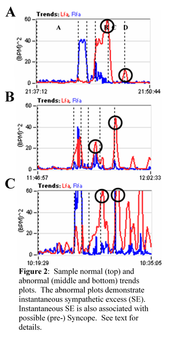
What is expected from P&S testing is one or more of four possible P&S Dysfunctions that underlie the Dysautonomia typically associated with Oxidative Stress.


REFERENCES

_________________

[1] Murray GL.  COVID-19 cardiac complications: Is an easy, safe treatment strategy right under our noses?  J Cardiovasc Dis Diag. 2020; 8:5.  doi: 10.37421/jcdd.2020.8.415.

2 DePace NL, Colombo J.  Autonomic and Mitochondrial Dysfunction in Clinical Diseases:  Diagnostic, Prevention, and Therapy.  Springer Science + Business Media, New York, NY, 2019.

3 Acosta C, DePace NL, DePace NL, Kaczmarski K, Pinales JM, and Colombo J.  Antioxidants effect changes in systemic parasympathetic and sympathetic nervous system responses and improve outcomes.  Cardio Open. 2020; 5(1): 26-36.  doi:  10.33140/COA.05.01.04

4 Colombo J, Arora RR, DePace NL, Vinik AI.  Clinical Autonomic Dysfunction:  Measurement, Indications, Therapies, and Outcomes.  Springer Science + Business Media, New York, NY, 2014.

5 Vinik A, Ziegler D.  Diabetic cardiovascular autonomic neuropathy.  Circulation. 2007; 115: 387-397.

6 Vinik AI, Maser RE, Nakave AA.  Diabetic cardiovascular autonomic nerve dysfunction.  US Endocrine Disease.  2007; Dec: 2-9.

7 Malik, M.  The Task Force of the European Society of Cardiology and the North American Society of Pacing and Electrophysiology. Heart rate variability, standards of measurement, physiological interpretation, and clinical use.  Circulation. 1996; 93:1043-1065.

8 Malik, M. and the Task Force of the European Society of Cardiology and the North American Society of Pacing and Electrophysiology. Heart rate variability, standards of measurement, physiological interpretation, and clinical use.  European Heart Journal. 1996, 17: 354-381.

9 Akselrod S, Oz O, Greenberg M, Keselbrener L.  Autonomic response to change of posture among normal and mild-hypertensive adults: investigation by time-dependent spectral analysis.  J Auton Nerv Syst. 1997 May 12;64(1):33-43.

10 Piña IL, Di Palo KE, Ventura HO.  Psychopharmacology and Cardiovascular Disease.  JACC. 2018; 71(20): 2346-2359.

11 Arora RR, Bulgarelli RJ, Ghosh-Dastidar S, Colombo J.  Autonomic mechanisms and therapeutic implications of postural diabetic cardiovascular abnormalities.  J Diabetes Science and Technology.  2008; 2(4): 568-71.

12 DePace NL, Vinik AI, Acosta C and Colombo J.  Oral vasoactive medications:  A Review of Midodrine, Droxidopa, and Pseudoephedrine as Applied to Orthostatic Dysfunction.  NEJM.  2020.  Submitted.

13 Vinik AI, Bloom HL, Colombo J.  Differential effects of adrenergic antagonists (carvedilol vs. metoprolol) on parasympathetic and sympathetic activity:  A comparison of measures.  Heart International. Heart Int. 2014; 9(1): 7-14; DOI: 10.5301/HEART.2014.12495.

14 Bloom HL, Vinik AI, Colombo J.  Differential effects of adrenergic antagonists (carvedilol vs. metoprolol) on parasympathetic and sympathetic activity:  A comparison of clinical results.  Heart Int. 2014 ; 9 (1): 15-21; DOI: 10.5301/HEART.2014.12496.

15 Murray GL and Colombo J.  (R)Alpha Lipoic Acid is a Safe, Effective Pharmacologic Therapy of Chronic Orthostatic Hypotension Associated with Low Sympathetic Tone.  Int J Angiol. In Print, 2018.


KEY WORDS

Coronavirus, Parasympathetic, Sympathetic, Oxidative Stress, Antioxidants


ABBREVIATIONS

ALA                            Alpha-Lipoic Acid

ANS                            Autonomic Nervous System

CoQ10                        Co-enzyme Q10

COVID-19                  Coronavirus (SARS-CoV-2)

P&S                             Parasympathetic and Sympathetic

PE                               Parasympathetic Excess

POTS                          Postural Orthostatic Tachycardia Syndrome

SE                               Sympathetic Excess

SW                              Sympathetic Withdrawal

[i] Murray GL.  COVID-19 cardiac complications: Is an easy, safe treatment strategy right under our noses?  J Cardiovasc Dis Diag. 2020; 8:5.  doi: 10.37421/jcdd.2020.8.415.

[ii] DePace NL, Colombo J.  Autonomic and Mitochondrial Dysfunction in Clinical Diseases:  Diagnostic, Prevention, and Therapy.  Springer Science + Business Media, New York, NY, 2019.

[iii] Acosta C, DePace NL, DePace NL, Kaczmarski K, Pinales JM, and Colombo J.  Antioxidants effect changes in systemic parasympathetic and sympathetic nervous system responses and improve outcomes.  Cardio Open. 2020; 5(1): 26-36.  doi:  10.33140/COA.05.01.04

Read More

Coronavirus Induces Oxidative Stress Leading to Autonomic Dysfunction – Part 1

Click here to download this post

Notes: This is the first in a series of 5 blog posts about COVID-19 and Autonomic Dysfunction.  This a pre-publication release that will be featured in a major medical journal.

Coronavirus Induces Oxidative Stress Leading to Autonomic Dysfunction Often With Delayed Symptom Onset

Heather L. Bloom, MD1 and Joseph Colombo, PhD, DNM, DHS2

  1. Electrophysiology, Atlanta Veterans Affairs Medical Center and Emory University Medical School, Atlanta, GA 
  2. Parasympathetic & Sympathetic Nervous System Consultant, Franklin Cardiovascular Associates, PA & Autonomic Dysfunction and POTS Center, Sewell, NJ, and Senior Medical Director & CTO, Physio PS, Inc., Atlanta, GA, dovetech@erols.com

Correspondence should be addressed to Dr. Colombo, dovetech@erols.com


ABSTRACT

Coronavirus, like other viruses and many chronic and serious pathologies, induce Oxidative Stress.  Oxidative Stress largely affects the Mitochondria of cells.  Cardiac and Nerve Cells are known to contain the largest numbers of Mitochondria of the cells in the body. 

The effect of Oxidative Stress on the Parasympathetic and Sympathetic (P&S) branches of the Autonomic Nervous System is to induce dysfunction.  P&S Dysfunction further affects the heart and other organs and systems of the body. 

Since the P&S branches are designed to work together to maintain normal organ function, even when dysfunctional, organ dysfunction is often delayed until P&S dysfunction is very significant.  Symptoms are not induced or realized until organ dysfunction presents. 

This delay in symptoms often appears to be healthy, and therefore is often not associated with the causal factor, such as Coronavirus.  Symptoms resulting from P&S dysfunction are often long-term and significantly impact patient quality of life and productivity. 

Symptoms include severe fatigue, anxiety, depression, lightheadedness, sleep difficulties, brain fog, cognitive and memory difficulties, GI disturbances, shortness of breath, palpitations, and more. 

Unfortunately, these symptoms are often not believed because these patients’ resting state is normal, including office exams, blood work, urine analysis, and other tests administered at rest, including many autonomic tests. 

However, the underlying P&S is not evident during the resting state it is only evident during the dynamic states when tests are typically not performed.  This editorial highlights the possibility of delayed Coronavirus-induced Oxidative Stress-induced P&S dysfunction, describes the possible types of P&S dysfunction, and offers possible therapy options to restore proper P&S function (balance).

ABBREVIATIONS

ALA                            Alpha-Lipoic Acid

ANS                            Autonomic Nervous System

CoQ10                        Co-enzyme Q10

COVID-19                  Coronavirus (SARS-CoV-2)

P&S                             Parasympathetic and Sympathetic

PE                               Parasympathetic Excess

POTS                          Postural Orthostatic Tachycardia Syndrome

SE                               Sympathetic Excess

SW                              Sympathetic Withdrawal

Read More