More Than Sick of Salt

All Posts Tagged: COVID-19

Chronic Fatigue Syndrome 101

What is Chronic Fatigue Syndrome (CFS) 101

Click here to download this post

Dr. DePace, MD, FACC

Symptoms of Chronic Fatigue Syndrome

Chronic diseases usually last for over 6 months. In Chronic Fatigue, we see post-exertional fatigue, unrefreshing sleep, and “Brain Fog” ( memory and cognitive disturbances).

The Autonomic Nervous System (Parasympathetic and Sympathetic balance) is often abnormal in Chronic Fatigue Syndrome (CFS). This affects blood pressure (BP) and heart rate (HR) regulation.

That’s why we see Orthostatic Intolerance in most cases like Postural Orthostatic Tachycardia Syndrome (POTS). In this condition they experience worsening symptoms when are in an upright position and improve when they lie down. Females are more affected than males. As many as 8 million Americans may be affected. Patients are affected at different ages and have different presentations.

What Causes Chronic Fatigue Syndrome?

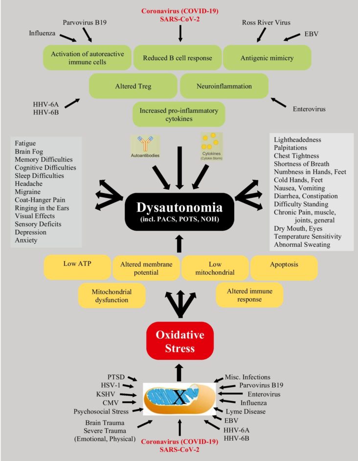
Chronic Fatigue Syndrome is a group of disorders that consists of many different causes. Let’s take them separately;

  1. Infection (viral or bacterial) that causes autoantibodies and oxidative stress to dysregulate cellular and specifically mitochondrial energetics. This may lead to exercise intolerance.
  2. Disturbed gut microbiota (abnormal bacterial colonization) possibly leading to “leaky-gut” leads to autoimmunity. Irritable Bowel Syndrome is seen in many Chronic Fatigue patients.
  3. Microglial activation of the nervous system, including the Central nervous System (CNS), possibly leading to chronic pain due to allodynia (pain due to stimuli that is usually not painful) and hyperalgesia (abnormally heightened sensitivity to pain).
  4. Neuronal inflammation is important in the pathophysiology of many disabling symptoms.
  5. High levels of pro inflammatory cytokines (chemicals produced by cells) and low level of antioxidants, such as Coenzyme Q-10 (CoQ10) or Glutathione
  6. Abnormalities of the Hypothalamus-Pituitary-Adrenal Axis possibly leading to “delayed cortisol awakening”, possibly leading to unrefreshing sleep. In some cases we see low cortisol levels. Cortisol is a hormone that helps the body handle stress.
  7. Physical or emotional trauma, including form an accident, concussion, immobilization, surgery, trauma, or even emotional stress such as loss of a loved one.
  8. Genetics may contribute, with identical twins having a higher incidence then fraternal twins. There has also been familial aggregations note of CFS.
  9. Environmental factors like mold or toxins may also be a triggers.

While mitochondrial dysfunction is implicated as an immediate cause of CFS, it is not determined what the damage to mitochondrial function is from.

Mitochondria are components of cells that are called organelles and they produce energy in the form of a molecule called ATP. Cellular hypoxia and oxidative stress happen during stressful situations.

Treatments For Chronic Fatigue Syndrome

The end result is disturbing muscle and nerve function. Exercise is the hallmark treatment for improving CFS. “Low and slow” exercise is where patients exercise 2-5 minutes followed by 5 minutes of rest so as not to damage skeletal muscle. Another such exercise is walking slowly, no more than 2 MPH for 40 minutes daily.

Even if biking or rowing, no more than 2 MPH. This may be too stressful for some patients, who on some days cannot lift their heads off the pillow. Supine exercises can be used for them. More work is required to assess the types of exercise programs that are most effective.

Diets high in processed foods and full of chemicals may be a cause of CFS and should be avoided. Cocktails of antioxidants that work on the mitochondria and immune system modulation are current areas of investigation. Currently, we are working on ways to categorize the different patients to determine which treatments work best.

Read More

Autonomic Nervous System and EDS (Ehlers-Danlos Syndrome)

Click here to download this post

Dr. DePace, MD, FACC

49% of hypermobile EDS (Ehlers-Danlos Syndrome) patients have POTS (postural orthostatic tachycardia syndrome), 31% orthostatic intolerance and 20% have normal hemodynamics. We call this orthostatic intolerance and postural orthostatic intolerance  in joint hypermobility syndrome / Ehlers-Danlos  hypermobility type, neurovegetative dysregulation or autonomic failure. The autonomic dysregulation is moderate to severe in one-third of our hypermobile EDS patients.

Coat-hanger pain is common in orthostatic intolerance associated with EDS. Coat-hanger syndrome consists of pain at the back of the neck (paracervical) and base of the head (suboccipital) that worsens in the upright position. It is believed to be due to poor blood flow to the muscles of the upper back and neck. It is due to pooling of blood due to abnormal sympathetic nervous system response due to standing and abnormal vasoconstriction. Coat-hanger pain can be quite profound, especially in conjunction with all the other chronic pain seen in EDS patients due to joint hypermobility.

Orthostatic headaches are also seen in EDS due to blood vessel malformation called Chiari malformation, CSF (cerebral spinal fluid) flow issues and CSF leaks. This may collagen problems, leading to stretchy blood vessels in EDS leading to venous pooling. This hypothesis has not been proven; however it makes empiric sense.

Autonomic dysfunction has often been attributed to autoimmunity and many times autoimmune antibodies are not detected, and many believe that this is because they have not been discovered as of yet. Diseases like rheumatoid arthritis, lupus and Sjogren’s disease have been seen with EDS. Nearly 10% had Raynaud’s, which is often associated with autoimmune disorders. It is kissable that abnormalities in the extracellular matrix might contribute to development of autoimmunity in the presence of other genetic or environmental influences.

The most common autoimmune diseases associated with EDS and POTS are Hashimoto’s, Sjogren’s, lupus and celiac disease. However, POTS is not the only dysautonomia disorder that is seen in EDS patients.

Mast cell activation syndrome is often seen in patients who have autonomic dysfunction including POTS and EDS. POTS and mast cell activation syndrome may frequently overlap. POTS patients with EDS tend to report dealing with POTS-like symptoms for most of their life. GI (gastrointestinal) are reported significantly more often by patients with EDS. Sensory neuropathic symptoms have been reported significantly more often in patients with EDS with POTS, including skin burning, hand tingling, hand burning, hand numbness and cold hands. The neuropathy noted in EDS patients suggests that the collagen in and around the nerve fibers may be damaged or abnormal.

Small fiber neuropathy in hypermobile EDS patients likely cause the burning sensations, hypesthesia and allodynia. Small fiber neuropathy refers to dysfunction or damage to the A-Delta and C fibers which relay thermal and nociceptive or unpleasant information as well as mediating autonomic function. There is strong evidence for a peripheral neuropathic contribution to the pain syndrome in hypermobile disorders in addition to the known nociceptive and central sensitization components. This raises the question if there is a neurological cause of hypermobile EDS; the only EDS syndrome without a known genetic cause. Physicians should assess for small fiber pathology in hypermobile EDS patients and hypermobility spectrum disorder patients for sensory and autonomic impact. EDS patients show an overactivity of the resting parasympathetic nerve tone and a decreased sympathetic nerve reactivity to stimuli.


Read More
DePace Books Heart Repair Manual, Clinical and Mitochondrial Disorders

Autonomic Nervous System Dysfunction in Ehlers Danlos Syndrome

Click here to download this post

Dr. DePace, MD, FACC

Articulo-autonomic dysplasia is a unifying pathogenic mechanism in Ehlers-Danlos Syndrome (EDS) and related conditions in the clinical pattern termed arthritis-adrenalin disorder.

Increased neuromuscular symptoms in females may be related to surrounding muscle support and joint connective tissue in males, leading to decreased male severity.

The similar clinical profiles of joint, skeletal, and dysautonomia, regardless of age our EDS diagnosis suggests the operation of an articulo-autonomic dysplasia (AAD) cycle, where lax vessels and lower body pooling elicit a sympathetic response and autonomic imbalance, which in turn affect small nerve fibers and enhance connective tissue laxity.

The findings of AAD are more frequent in females but are paralleled by men.

These include undergoing back surgery, slow healing, bladder issues, hernias, valve regurgitation, gallbladder issues, hives and reactive skin, food and medicine intolerance, and hypothyroidism.

The greater flexibility and fragility of connective tissue in women is conveyed by measures ranging from the historical performance of hypermobility tricks to the physical performance of Beighton maneuvers.

Neuromuscular and dysautonomia symptoms more frequent in women include migraines, muscle aches, weakness, and atrophy, physically highlighting greater muscular development and support in men as a key factor in their reduced severity.

The role of surrounding muscle for joint connective tissue constraint and protection correlates with the benefits of physical therapy and exercise for the treatment of EDS.

Problems can include popping joints that may manifest as subluxations, polyarticular and symmetrical joint pain of knees, shoulders, and ankles with rare swelling and erythema, joint injuries in mostly ankles and knees, fractures most frequently in distal limbs, and disk degeneration or herniation.

Clumsiness from joint laxity, cumulative joint pain injury, and skeletal deformations like scoliosis, toeing in or out, and flat or high arches make the typical patient uncomfortable with sports and prone to inactivity.

Evidence of skin fragility is another hallmark of EDS with easy bruising, unusual scars, and early striae.

Most unnoticed unless questioned or documented is soft or elastic skin which can be pulled in 1-inch folds from their jaw liner mid-forearm on physical examination.

The common findings of migraines and daily headaches which may arise from blood vessel abnormalities like Chiari formation of crania-cervical stability leading to numbness, tingling and muscle aches can prompt fibromyalgia diagnosis.

Seizures may actually reflect syncope more than epilepsy and may be related to poor balance.

Bloating, stomach pain and nausea begin early in life and continue later with gall bladder dysfunction and are accentuated by mast cell activation disease that presents as eosinophilic esophagitis with frequent food intolerances.

It is common to misdiagnose the anxiety and tachycardia associated with Postural tachycardia syndrome (POTS) as a functional disease.

The bowel disorders and overlapping joint and autonomic symptoms seen in EDS often are confused with Crohn’s and Celiac disease with the various associated psychological aspects.

Genomic and immunological studies can help determine if the overlapping joint and autonomic symptoms have separate causes. It is also important to make sure there is not a vitamin D deficiency or hypothyroidism that can present as Hashimoto’s thyroiditis.

In addition to chronic fatigue, anxiety-tachycardia and POTS, we see brain fog – poor focus, and sleep disturbances.

This can be disabling and much more severe in females with the occasional extremely affected male and together with bowel issues, weight loss, hives and reactive skin, and reactive airway disease – shortness of breath.

Mild valvular regurgitation, mostly mitral prolapse in both sexes.

Findings related to Marfanoid habits include an angular build, arm span greater than height, and long fingers with consequent maneuvers like making the prayer sign behind the back. Deformations like neck kyphosis, scoliosis, and lordosis are much more frequent in females except for pectus and toeing-in mainly or out, a likely contributor to clumsiness.

Read More

Long-COVID Syndrome and the Cardiovascular System: A Review of Neurocardiologic Effects on Multiple Systems

Click here to download this post

Dr. DePace, MD, FACC

Dr. Joseph Columbo


Note: This is an article recently published by Springer in their “Current Cardiology Reports” on their website. The Abstract is printed here.  The entire article is availalbe by clicking this link or downloading it as a PDF 

Abstract

Purpose of Review

Long-COVID syndrome is a multi-organ disorder that persists beyond 12 weeks post-acute SARS-CoV-2 infection (COVID-19). Here, we provide a definition for this syndrome and discuss neuro-cardiology involvement due to the effects of (1) angiotensin-converting enzyme 2 receptors (the entry points for the virus), (2) inflammation, and (3) oxidative stress (the resultant effects of the virus).

Recent Findings

These effects may produce a spectrum of cardio-neuro effects (e.g., myocardial injury, primary arrhythmia, and cardiac symptoms due to autonomic dysfunction) which may affect all systems of the body. We discuss the symptoms and suggest therapies that target the underlying autonomic dysfunction to relieve the symptoms rather than merely treating symptoms. In addition to treating the autonomic dysfunction, the therapy also treats chronic inflammation and oxidative stress. Together with a full noninvasive cardiac workup, a full assessment of the autonomic nervous system, specifying parasympathetic and sympathetic (P&S) activity, both at rest and in response to challenges, is recommended. Cardiac symptoms must be treated directly. Cardiac treatment is often facilitated by treating the P&S dysfunction. Cardiac symptoms of dyspnea, chest pain, and palpitations, for example, need to be assessed objectively to differentiate cardiac from neural (autonomic) etiology.

Summary

Long-term myocardial injury commonly involves P&S dysfunction. P&S assessment usually connects symptoms of Long-COVID to the documented autonomic dysfunction(s).

Read More
Ehlers-Danlos Syndrome

Vascular Ehlers-Danlos Syndrome Part 2

Click here to download this post

Dr. DePace, MD, FACC


Note: This Post is a contuinuation of the September 13, 2022 post on Vascular EDS

EDS has different types. Type IV is known as Vascular EDS. Pregnancy increases the likelihood of uterine or vascular rupture in women suffering from EDS type IV particularly during the last 3 months. The highest risk of rupture is during labor. Uterine hemorrhage can also occur postpartum. Occasionally a hysterectomy has been needed. The value of a Cesarean section carried out before the onset of labor has not been proven. Some have proposed that a medication called desmopressin be used to control bleeding during delivery.

The tendency towards hemorrhage in Type IV EDS is due to the fragility of tissue and capillaries rather than something wrong with the blood. The problem is due a defect in the protein collagen that makes fibers in arterial and venous blood vessels. This defect is what accounts for tears or dissections. This is one reason why digestive perforations can occur frequently.

Pregnant women with vascular EDS should receive treatment at special clinics. Because it is genetic, once EDS type IV is identified, Genetic counseling is recommended. The transmission is autosomal dominant, which means you only need one parent to pass on the trait, however 50% can be spontaneous with no family history.

A conservative approach in the management of EDS type IV is usually recommended. Avoid intense physical activities, violent sports, contact sports and scuba diving. Avoid drugs that interfere with platelet function or coagulation, like anticoagulants or vitamin K antagonist. Arteriograms and endoscopies are usually relatively contraindicated in GI and uterine complications unless absolutely necessary.

Surgery may, however, may be required urgently to treat potentially fatal complications, especially with very large or expanding aneurisms, or in the case of dissections and in the case of hemorrhage. Special techniques need to be used and information on the use of stents to treat vascular complications of EDS type IV is insufficient. That being said, simple arterial repairs have been successfully carried out.

Read More