More Than Sick of Salt

All Posts in Category: parasympathetic nervous system

Autonomic Nervous System and EDS (Ehlers-Danlos Syndrome)

Click here to download this post

Dr. DePace, MD, FACC

49% of hypermobile EDS (Ehlers-Danlos Syndrome) patients have POTS (postural orthostatic tachycardia syndrome), 31% orthostatic intolerance and 20% have normal hemodynamics. We call this orthostatic intolerance and postural orthostatic intolerance  in joint hypermobility syndrome / Ehlers-Danlos  hypermobility type, neurovegetative dysregulation or autonomic failure. The autonomic dysregulation is moderate to severe in one-third of our hypermobile EDS patients.

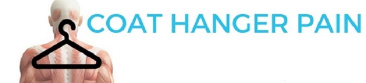
Coat-hanger pain is common in orthostatic intolerance associated with EDS. Coat-hanger syndrome consists of pain at the back of the neck (paracervical) and base of the head (suboccipital) that worsens in the upright position. It is believed to be due to poor blood flow to the muscles of the upper back and neck. It is due to pooling of blood due to abnormal sympathetic nervous system response due to standing and abnormal vasoconstriction. Coat-hanger pain can be quite profound, especially in conjunction with all the other chronic pain seen in EDS patients due to joint hypermobility.

Orthostatic headaches are also seen in EDS due to blood vessel malformation called Chiari malformation, CSF (cerebral spinal fluid) flow issues and CSF leaks. This may collagen problems, leading to stretchy blood vessels in EDS leading to venous pooling. This hypothesis has not been proven; however it makes empiric sense.

Autonomic dysfunction has often been attributed to autoimmunity and many times autoimmune antibodies are not detected, and many believe that this is because they have not been discovered as of yet. Diseases like rheumatoid arthritis, lupus and Sjogren’s disease have been seen with EDS. Nearly 10% had Raynaud’s, which is often associated with autoimmune disorders. It is kissable that abnormalities in the extracellular matrix might contribute to development of autoimmunity in the presence of other genetic or environmental influences.

The most common autoimmune diseases associated with EDS and POTS are Hashimoto’s, Sjogren’s, lupus and celiac disease. However, POTS is not the only dysautonomia disorder that is seen in EDS patients.

Mast cell activation syndrome is often seen in patients who have autonomic dysfunction including POTS and EDS. POTS and mast cell activation syndrome may frequently overlap. POTS patients with EDS tend to report dealing with POTS-like symptoms for most of their life. GI (gastrointestinal) are reported significantly more often by patients with EDS. Sensory neuropathic symptoms have been reported significantly more often in patients with EDS with POTS, including skin burning, hand tingling, hand burning, hand numbness and cold hands. The neuropathy noted in EDS patients suggests that the collagen in and around the nerve fibers may be damaged or abnormal.

Small fiber neuropathy in hypermobile EDS patients likely cause the burning sensations, hypesthesia and allodynia. Small fiber neuropathy refers to dysfunction or damage to the A-Delta and C fibers which relay thermal and nociceptive or unpleasant information as well as mediating autonomic function. There is strong evidence for a peripheral neuropathic contribution to the pain syndrome in hypermobile disorders in addition to the known nociceptive and central sensitization components. This raises the question if there is a neurological cause of hypermobile EDS; the only EDS syndrome without a known genetic cause. Physicians should assess for small fiber pathology in hypermobile EDS patients and hypermobility spectrum disorder patients for sensory and autonomic impact. EDS patients show an overactivity of the resting parasympathetic nerve tone and a decreased sympathetic nerve reactivity to stimuli.


Read More
DePace Books Heart Repair Manual, Clinical and Mitochondrial Disorders

Autonomic Nervous System Dysfunction in Ehlers Danlos Syndrome

Click here to download this post

Dr. DePace, MD, FACC

Articulo-autonomic dysplasia is a unifying pathogenic mechanism in Ehlers-Danlos Syndrome (EDS) and related conditions in the clinical pattern termed arthritis-adrenalin disorder.

Increased neuromuscular symptoms in females may be related to surrounding muscle support and joint connective tissue in males, leading to decreased male severity.

The similar clinical profiles of joint, skeletal, and dysautonomia, regardless of age our EDS diagnosis suggests the operation of an articulo-autonomic dysplasia (AAD) cycle, where lax vessels and lower body pooling elicit a sympathetic response and autonomic imbalance, which in turn affect small nerve fibers and enhance connective tissue laxity.

The findings of AAD are more frequent in females but are paralleled by men.

These include undergoing back surgery, slow healing, bladder issues, hernias, valve regurgitation, gallbladder issues, hives and reactive skin, food and medicine intolerance, and hypothyroidism.

The greater flexibility and fragility of connective tissue in women is conveyed by measures ranging from the historical performance of hypermobility tricks to the physical performance of Beighton maneuvers.

Neuromuscular and dysautonomia symptoms more frequent in women include migraines, muscle aches, weakness, and atrophy, physically highlighting greater muscular development and support in men as a key factor in their reduced severity.

The role of surrounding muscle for joint connective tissue constraint and protection correlates with the benefits of physical therapy and exercise for the treatment of EDS.

Problems can include popping joints that may manifest as subluxations, polyarticular and symmetrical joint pain of knees, shoulders, and ankles with rare swelling and erythema, joint injuries in mostly ankles and knees, fractures most frequently in distal limbs, and disk degeneration or herniation.

Clumsiness from joint laxity, cumulative joint pain injury, and skeletal deformations like scoliosis, toeing in or out, and flat or high arches make the typical patient uncomfortable with sports and prone to inactivity.

Evidence of skin fragility is another hallmark of EDS with easy bruising, unusual scars, and early striae.

Most unnoticed unless questioned or documented is soft or elastic skin which can be pulled in 1-inch folds from their jaw liner mid-forearm on physical examination.

The common findings of migraines and daily headaches which may arise from blood vessel abnormalities like Chiari formation of crania-cervical stability leading to numbness, tingling and muscle aches can prompt fibromyalgia diagnosis.

Seizures may actually reflect syncope more than epilepsy and may be related to poor balance.

Bloating, stomach pain and nausea begin early in life and continue later with gall bladder dysfunction and are accentuated by mast cell activation disease that presents as eosinophilic esophagitis with frequent food intolerances.

It is common to misdiagnose the anxiety and tachycardia associated with Postural tachycardia syndrome (POTS) as a functional disease.

The bowel disorders and overlapping joint and autonomic symptoms seen in EDS often are confused with Crohn’s and Celiac disease with the various associated psychological aspects.

Genomic and immunological studies can help determine if the overlapping joint and autonomic symptoms have separate causes. It is also important to make sure there is not a vitamin D deficiency or hypothyroidism that can present as Hashimoto’s thyroiditis.

In addition to chronic fatigue, anxiety-tachycardia and POTS, we see brain fog – poor focus, and sleep disturbances.

This can be disabling and much more severe in females with the occasional extremely affected male and together with bowel issues, weight loss, hives and reactive skin, and reactive airway disease – shortness of breath.

Mild valvular regurgitation, mostly mitral prolapse in both sexes.

Findings related to Marfanoid habits include an angular build, arm span greater than height, and long fingers with consequent maneuvers like making the prayer sign behind the back. Deformations like neck kyphosis, scoliosis, and lordosis are much more frequent in females except for pectus and toeing-in mainly or out, a likely contributor to clumsiness.

Read More
Ehlers-Danlos Syndrome

Vascular Ehlers-Danlos Syndrome Part 2

Click here to download this post

Dr. DePace, MD, FACC


Note: This Post is a contuinuation of the September 13, 2022 post on Vascular EDS

EDS has different types. Type IV is known as Vascular EDS. Pregnancy increases the likelihood of uterine or vascular rupture in women suffering from EDS type IV particularly during the last 3 months. The highest risk of rupture is during labor. Uterine hemorrhage can also occur postpartum. Occasionally a hysterectomy has been needed. The value of a Cesarean section carried out before the onset of labor has not been proven. Some have proposed that a medication called desmopressin be used to control bleeding during delivery.

The tendency towards hemorrhage in Type IV EDS is due to the fragility of tissue and capillaries rather than something wrong with the blood. The problem is due a defect in the protein collagen that makes fibers in arterial and venous blood vessels. This defect is what accounts for tears or dissections. This is one reason why digestive perforations can occur frequently.

Pregnant women with vascular EDS should receive treatment at special clinics. Because it is genetic, once EDS type IV is identified, Genetic counseling is recommended. The transmission is autosomal dominant, which means you only need one parent to pass on the trait, however 50% can be spontaneous with no family history.

A conservative approach in the management of EDS type IV is usually recommended. Avoid intense physical activities, violent sports, contact sports and scuba diving. Avoid drugs that interfere with platelet function or coagulation, like anticoagulants or vitamin K antagonist. Arteriograms and endoscopies are usually relatively contraindicated in GI and uterine complications unless absolutely necessary.

Surgery may, however, may be required urgently to treat potentially fatal complications, especially with very large or expanding aneurisms, or in the case of dissections and in the case of hemorrhage. Special techniques need to be used and information on the use of stents to treat vascular complications of EDS type IV is insufficient. That being said, simple arterial repairs have been successfully carried out.

Read More
Vascular EDS

Vascular Ehlers-Danlos Syndrome

Click here to download this post

Dr. DePace, MD, FACC

Ehlers-Danlos Syndrome, Vascular Type

The vascular type of Ehlers-Danlos syndrome is predisposed to blood vessel and bowel rupture. Vascular EDS is not as common as hypermobile EDS, but important to recognize because of vascular complications.

Physical Characteristic Vascular Ehlers-Danlos Syndrome

The main presentation is hematoma (bruise or collection of blood) in a muscle. More serious and less common would be intracranial hemorrhage (bleeding in the skull).

Another serious complication, even less common, is arterial dissections (splitting of the wall of the blood vessel. Vascular EDS is not the only subtype of EDS  that presents with vascular complications. Hypermobile EDS is more common but rarely has vascular complications.

About 2% of non-vascular EDS have vascular complications.  The most common such problems include hematoma, then intracranial hemorrhage, arterial dissection, arterial aneurism,  GI bleeding, and operative hemorrhage or sporadic vascular complications.

In addition, venous complications such as varicose veins and deep vein thrombosis were reported. Referral for cardiovascular assessment and regular follow-up may be required.

Treatment for Vascular Ehlers-Danlos Syndrome

Therapeutic measures are limited to the treatment of symptoms in vascular EDS. There is only one evidence  based preventative medication called  Celiprolol, which reduces heart rate and pulsatile pressures if there is high blood pressure and can decrease continuous and pulsatile mechanical stress on collagen fiber within the arterial wall.

It should be emphasized that certain genetic determinants result in a shorter life expectancy. The goal is to delay the onset of complications.

Read More

Classical Type Ehlers-Danlos Syndrome

Click here to download this post

Dr. DePace, MD, FACC


Within the system of Classical Ehlers-Danlos Syndrome (EDS), there were initially 6 subtypes. However, with the identification of additional genetic and biochemical markers, in 2017 the classification was revised to include 13 types. We previously discussed vascular EDS  in greater detail   in the last blog. In this blog, we will discuss classical EDS.

With classical EDS, the major diagnostic criteria include skin hyperextensibility, widened atrophic scars, and joint hypermobility. It should be noted that the severity varies even among family members.

Certain proteins called collagen, which provide strength and structure to the extracellular matrix of tissues and organs. The genes that are associated with classical EDS are passed on with autosomal dominant inheritance, which means you only need one parent to get the gene.

Classical Type Ehlers-Danlos Syndrome Criterion

The Major criteria include:

  1. Skin hyperextensibility and atrophic scarring.
  2. General hypermobility (a Beighton score of 5 or more)

Minor criteria include:

  1. Easy bruising
  2. Soft doughy skin
  3. Skin Fragility or traumatic splitting
  4. Mulluscoid pseudotumors, which are fleshy lesions associated with scars at pressure points.
  5. Subcutaneous spheroids, which are small round hard bodies that are mobile and commonly located on the forearms and chin.
  6. Hernia or history of hernia
  7. Epicanthal folds
  8. Complications of joint hypermobility such as sprains, subluxation, pain, or flexible flat foot.
  9. First degree relatives who meet clinical criteria.


Minimal criteria suggestive of classical EDS include skin hyperextensibility and atrophic scaring plus generalized joint hypermobility and 3 or more minor criteria.

Genetic confirmation is required for a definitive diagnosis. More than 90% of classical EDS patients labor a mutation of one of the genes that encode for type V collagen. A reduction in type V collagen is central to the pathogenesis of classical EDS.

While musculoskeletal joint hyper-mobility is present in classical EDS, the skin is the key to establishing the diagnosis of classical EDS. The skin is hyperextensible and soft with severe atrophic scarring and hemosiderin deposits, or brown areas over the shin and extensor surfaces, due to easy bruising. Poor wound healing is often seen in classical EDS.

A characteristic facial feature has been described. These are epicanthic folds, excess skin on the eyelids, a prematurely aged appearance, and scars on the forehead and chin. The absence of striae or stretch marks has also been noted in classical EDS patients.

Other problems are gastrointestinal problems, most commonly nausea, vomiting, and gastroesophageal reflux, followed by chronic constipation. Abnormalities in the cornea are also found in classical EDS with thin and steep and transparent corneas.

Read More