More Than Sick of Salt

All posts by joeobiso

Long COVID Symptoms

Click here to download this post

By Dr. Nicholas DePace and Dr. Joseph Columbo

Long‑COVID Symptoms

Long-COVID symptoms may be explained by a pro-inflammatory state with oxidative stress and P&S dysfunction.

Cardiopulmonary testing for unexplained dyspnea post-COVID-19 was reported. Patients with symptoms consistent with chronic fatigue had an abnormal pattern of oxygen uptake on cardiopulmonary testing consistent with what is seen with chronic fatigue syndrome.

Circulatory impairment, abnormal ventilatory pattern, and chronic fatigue syndrome may be common in patients with post-acute sequelae of post-concussive syndrome, and this accounts for the mechanism of dyspnea in many patients who do not have pulmonary disease from Long-COVID or myocardial dysfunction.

The symptoms of Long-COVID syndrome may all be associated with autonomic dysfunction as measured with cardiorespiratory testing and Current Cardiology Reports 1 3 relieved with appropriate parasympathetic or sympathetic therapies based on the cardiorespiratory test.

This Post is an excerpt from Current Cardiology Reports: https://link.springer.com/article/10.1007/s11886-022-01786-2

Read More

Defining Long COVID

Click here to download this post

By Dr. Nicholas DePace and Dr. Joseph Columbo

Long‑COVID Definition

What exactly is Long-COVID syndrome?

Long-COVID or post-COVID-19 is an umbrella term that refers to symptoms persisting past the initial phase. There are many definitions that have been offered.

Official Definition of Long-COVID

Long-COVID has recently been defined as “the condition that occurs in individuals with a history of probable or confirmed SARS-CoV-2 infection, usually 3 months from the onset of COVID-19, with symptoms that last for at least 2 months and cannot be explained by an alternative diagnosis”

 Likewise, there now exists an International Classification of Diseases, Tenth Revision (ICD-10) code corresponding to Long-COVID condition—U09.9.  Basically, there are individuals who do not completely recover over a period of weeks, usually 2–3 weeks. Since COVID-19 is a novel disease, there is still no consensus of the definition of Long-COVID symptoms.

Prevalence and Symptom Categories

Systematic Review Findings

A systematic review documented 20% of the reports of long-term COVID symptoms involved abnormal lung function, 24% involved neurological complaints and olfactory dysfunction, and 55% on specific widespread symptoms, mainly chronic fatigue and pain.

WHO’s Clinical Case Definition

The World Health Organization (WHO) developed a clinical case definition of Long-COVID by Delphi methodology that included 12 domains. However, the understanding of this definition has been going through changes as new evidence emerges, and we are gaining a better understanding of the consequences of COVID-19 and its mutations.

Symptom Duration and Impact

Usually, three or more months past the acute COVID-19 infection, symptoms that last for at least 2 months and cannot be explained by alternate diagnoses may ft this definition. These symptoms include fatigue, shortness of breath, cognitive dysfunction, and symptoms that affect the functional capacity of patients with daily living and productivity. Symptoms may fluctuate, fare up, or relapse over time, adversely affecting multiple organ systems.

P&S Nervous Systems Dysfunction in Long Covid

Role of P&S Nervous Systems

We propose that the delay between surviving the acute COVID infection and the onset of the Long-COVID symptoms is a function of the P&S nervous systems.

The P&S nervous systems function together to coordinate and control organs and organ systems to maintain normal organ function, even when the two nervous systems are dysfunctional.

Dysfunction and Symptoms

Prolonged P&S dysfunction, once severe enough, then leads to poor organ control and then symptoms. This process may take up to 3 months, faster if there were prior comorbidities, including age.

This is the basis for our claims that Long-COVID is a combination of both parasympathetic dysfunction(s) and sympathetic dysfunction(s). In our experience, the prolonged severe immune responses to COVID-19 seems to cause prolonged excessive parasympathetic responses, leading to secondary, prolonged, excessive beta-adrenergic (sympathetic) responses which prolongs and exaggerates heart rate, blood pressure, histaminergic, inflammatory, pain, and anxiety responses.

Gastrointestinal and Other Symptoms

The parasympathetic excess may also lead to both upper and lower GI symptoms.

The oxidative stress of the acute COVID-19 infection also causes oxidative stress which often leads to alpha-adrenergic (sympathetic) dysfunction which leads to orthostatic dysfunction and poor coronary and cerebral perfusion and the perfusion of the anatomy in between causing many of the rest of the symptoms of Long-COVID. Long-COVID may directly affect the lungs, heart, nervous system, kidneys, and pancreas.

Challenges in Research and Treatment

Lack of Standardized Definition

Unfortunately, the lack of a standardized definition for Long-COVID syndrome presents obstacles for researchers Fig. 1 The cytokine storm involved in COVID-19 infections is a source of oxidative stress.

Viruses and traumas (mental or physical) in general may lead to oxidative stress, which may lead to parasympathetic or sympathetic dysfunction(s), known as dysautonomia (adapted from Rasa et al.) ◂ Current Cardiology Reports 1 3 in studying the condition with controlled studies and arriving at a precise diagnosis and treatment algorithms. In addition, many patients with Long-COVID syndrome require rehospitalization especially those with comorbidities, such as cardiovascular disease, diabetes mellitus, obesity, cancer, and kidney disease.  


This Post is an excerpt from Current Cardiology Reports: https://link.springer.com/article/10.1007/s11886-022-01786-2

Read More

Parasympathetic and Sympathetic, Nervous System Dysfunction and Monitoring – Introduction

Click here to download this post

By Dr. Nicholas DePace and Dr. Joseph Columbo

There are many consequences of Parasympathetic and Sympathetic Dysfunction (P&S Dysfunction; aka., Autonomic Dysfunction or Dysautonomia) 

Major consequences and their effects include:

  • Abnormal cardiovascular control, causing inappropriate peripheral vasoconstriction, inappropriate shifts in blood volume, poor perfusion and distribution, and inefficient cardiac contractility.  The net effects are (1)gravitational pooling of blood volume, usually to the lower half of the body upon or during upright posture; (2) difficulty in returning blood to the heart to produce an adequate cardiac output, which results in inappropriate rapid drops in BP or rapid increases in HR; and (3) an inappropriate distribution of blood flow throughout the various organs of the body, some receiving too much and some too little, resulting in many of the symptoms listed near the end of this Introduction;
  • P&S imbalance, both at rest and in response to challenges, causing abnormal organ function even when the organs themselves are normal and healthy.  Examples include: (1) unequal pupil sizes; (2) abnormal motility and pH of the gastrointestinal (GI) tract; (3) hormone dysregulation; (4) abnormal pain, inflammatory, histaminergic, BP, HR, blood glucose, or affective (g., anxiety or depression, ADD/ADHD, OCD, PTSD) responses; and
  • Compensation, causing many adverse symptoms that may arise when the body attempts to compensate for the P or S dysfunction, such as high resting BP (possibly resulting in secondary hypertension) in response to Orthostatic or Syncopal disorders and auto-immune-like responses resulting from exaggerated inflammatory responses due to Sympathetic Excess secondary to Parasympathetic Excess (see below).
  • Note: In cases where the P&S dysfunctions are relieved and symptoms persist, then the remaining dysfunctions or disorders are true end-organ dysfunctions and should be treated accordingly.

Read More

Signs and Symptoms of Dysautonomia (Autonomic Dysfunction)

Click here to download this post

By Dr. Nicholas DePace and Dr. Michael Goldis

Symptoms of Dysautonomia include fainting. drop in blood pressure with getting up, blood pressures that occur without a known cause, flushing of the face, lack of sweating or sweating too much, vomiting, constipation, diarrhea, difficulty swallowing, swelling of the belly, disturbances of urination, difficulties with erections, episodes of stopping breathing and the declining ability to see at night. Quite a lot!

Before the person actually faints, they may experience loss of strength in the muscles that keep us upright, weakness, a generalized feeling of not being well, nausea, headache, sweating, a pale complexion, difficulty seeing and a sense that they are about to lose consciousness. 

Usually low blood pressure and slow heart beat accompany these symptoms. These signs and symptoms can be caused by emotional stress, drops in blood pressure when getting up, vigorous exercise in a hot environment, blockage of blood returning to the heart, sudden onset of pain or its anticipation, and loss of fluids.

There can be a variety of other circumstances involved with these feelings faint one gets before they faint due to dysautonomia.

When a person does faint, they usually limp. Some muscle movement may occur and they may experience fainting and sometimes lose bowel control which can appear like seizure. what is different is that recovery is rapid  once the person is lying flat. After the fainting event, headache, nausea and fatigue. usually persist.


Reference – Current Medical Diagnosis and Treatment, Dysautonomia  2021 page 101


Read More
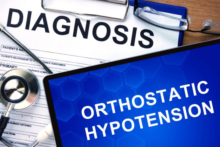
What is Orthostatic Hypotension

Cracking the Code of Dysautonomia: POTS, Orthostatic Hypotension, and Heart Health

Click here to download this post

by Nicholas DePace MD, FACC and Michael Edward Goldis DO, FACOI, MS, BS in Pharm

When venous pooling occurs, the cardiovascular system attempts to maintain cardiac output with activation of the sympathetic nervous system, the so called accelerator of the body.

Adrenalin is released in above normal amounts and that causes the heart to beat faster or compensatory tachycardia. In addition, increased vascular tone occurs with alpha agonist activity.

This maintains blood pressure while there is decreased preload from venous pooling and maintains cardiac output by increasing heart rate with a reduced stroke volume.

This explains how cerebral circulation and thus consciousness is maintained in compensated states like orthostatic hypotension and POTS.

The increased heart rate increased the output of the heart (the cardiac output) while the actual volume the heart puts out each stroke (the stroke volume) falls because of venous pooling.

The heart rate may increase 30, 40 or more above baseline. Therefore, POTS is known as a compensated neuro-cardiovascular dysfunction. One could argue that this is not a well-compensation as the patient has significant symptoms still resulting in orthostatic intolerance.

In contrast, Orthostatic hypotension is thought of as an uncompensated neuro-cardiovascular dysfunction. Here, the patient can become dizzy and lightheaded because the blood pressure is not maintained when there is venous pooling.

Because the heart rate generally does not increase significantly for compensation, the patient’s blood pressure drops and may even have overt syncope.

Patients with orthostatic hypotension often do not have an adequate rise in heart rate because of sympathetic nervous system decompensation.

A person can have a 30 or 40 point rise in heart rate, meeting the criteria for POTS, and in another moment, when the neuro-cardiovascular system is not compensated, they can have a blood pressure drop resulting in orthostatic hypotension.

So, while it is rare, both POTS and orthostatic hypotension can coexist. Generally, if someone demonstrates orthostatic hypotension, they rarely have POTS and vice versa.

Vasovagal syncope is a sort of form of orthostatic hypotension which is delayed.

This is whether is an increasing vagal tone that prevents blood vessels from adequate constriction and prevents the heart rate from adequately increasing.

This is an extremely delayed form of orthostatic hypotension which sometimes can be reproduced on Tilt Testing.

POTS oftentimes is due to several mechanisms. There can be hypovolemic, hyperadrenergic, and neuropathic POTS. Mast cell activation is a potential mechanism as well as certain enzyme deficiencies.

When the sympathetic nervous system is extremely overactive the blood pressure may even elevate with a rise in heart rate, which is hyperadrenergic POTS.

Regardless of the mechanism of POTS, the treatment is basically similar. But, for hypovolemic POTS a volume expander makes clinical sense and for hyperadrenergic POTS, a beta blocker makes sense.

Neuropathic POTS occurs when there are diseased small fibers, which can happen with diabetes, rheumatoid arthritis, lupus, and Sjogren’s syndrome and usually has some degree of autoimmunity, but may not necessarily occur in small fiber neuropathy.

While small fiber biopsy is the gold standard to diagnose this form of POTS, we have used psuedomotor testing as an alternative.

The end result, whether the dysautonomia is due to sympathetic overdrive or parasympathetic dysfunction is poor perfusion to the brain, leading to dizziness, syncope, vision and hearing loss, tinnitus, and brain fog.

With Sympathetic overdrive, which is a major compensation mechanism, the patient has a racing heart, insomnia, excessive sweating, high anxiety, and exacerbation of brain fog from reduced cerebral blood flow from hyperventilation. This may also explain the palpitations a person feels with dysautonomia.


Read More