More Than Sick of Salt

All Posts Tagged: orthostatic hypotension

Ehlers-Danlos Syndrome (EDS)- Part 1 – Introduction, What is it?

Click Here to Download this Blog Post –  Ehlers-Danlos Syndrome (EDS)- Part 1 – Introduction, What is it?

Dr. Nicholas L. DePace, M.D., F.A.C.C.

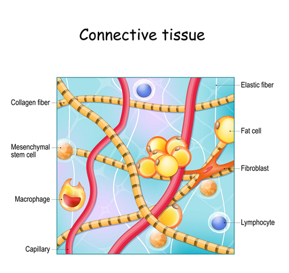
Ehlers-Danlos Syndrome (EDS), what is it? EDS comprises a group of more than 10 different inherited, clinically, and genetically diverse in character, disorders, resulting from genetic defects in collagen, a group of fibers proteins involved with connective-tissue synthesis and structure. The different variants of EDS due to biochemical and genetic differences is what creates the clinical differences. Many individuals do not fit into a single type  and overlap is common. Symptoms can include highly flexible, loose joints, joint pain, fragile and redundant skin with a velvety texture, and abnormal scar formation which can be seen at birth or in early childhood.

The most common forms of  EDS are not life threatening or dangerous. There are some rare types that can be. Some manifestations that are only seen in this rare types of EDS include:

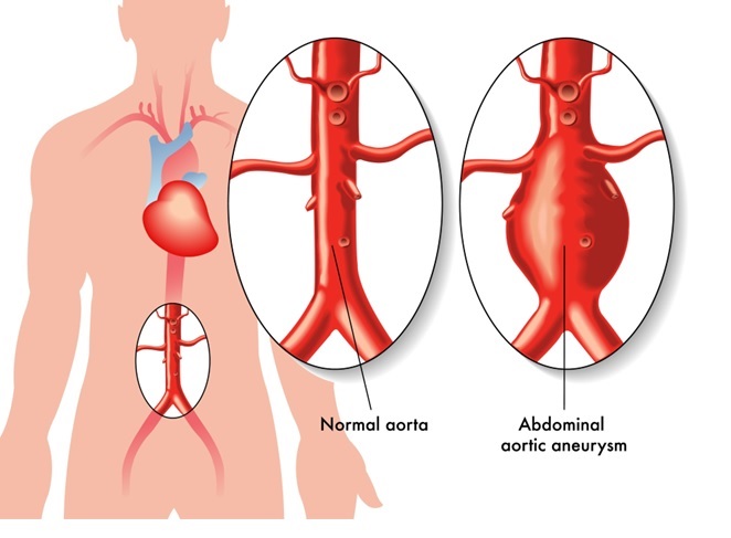
Aortic dissection, which is a medical emergency in which a tear happens in the inner layer of the body’s main artery, the aorta, can occur. Blood rushes through the tear causing the middle layers of the artery to split. This is called a dissection. If the blood goes outside the artery, it can be deadly.

Scoliosis, where the spine can curve to either side, can change a person’s posture. The shoulders aren’t even and can even affect breathing movement.

Brittle cornea is a rare but significant variant of EDS, which predisposes people to corneal ruptures and possible blindness.

Blue sclerae is when the white of the eye appears blue, great or even purple. The condition is usually painless and is due to thinning of the connective tissue proteins letting the blood vessels underneath show through. In itself, the condition is not dangerous, but means there is a significant underlying medical condition that will need treatment.

Muscles waste away.  Hypotonia is a state of reduced muscle strength.

Postural hypotension is when the blood pressure drops on changing to an upright position, also call Orthostatic Hypotension. It can cause dizziness or lightheadedness and possibly fainting.

Cardiac valvular abnormalities are conditions that can cause one or more heart valves to leak blood backward into the heart chambers or fail to open fully. Your doctor may hear a “whooshing” noise or an extra click with a stethoscope. Some people have no symptoms, but poorly functioning heart valves can cause chest pain, difficulty breathing, tiredness, weakness, feeling irregular heartbeats, lightheadedness, fainting and swollen ankles, feet or belly.

Chronic pain can also affect those afflicted by EDS.

It is important to seek out a clinician with expertise in EDS to make an accurate diagnosis and create a treatment plan. One of the nation’s leading centers is Franklin Cardiovascular Associates, under the direction of Nicholas DePace, MD, FACC. They are located in Sicklerville, New Jersey. franklincardiovascular.com, (856) 589-6034

Read More
Orthostatic Hypotension Blog 6 of 6

Mechanisms and Rare Causes of Neurogenic Orthostatic Hypotension (Part 6 Of 6)

Click Here to Download this Blog Post –  Orthostatic Hypotension: Part 6 Of 6

Dr. Nicholas L. DePace, M.D., F.A.C.C.

This is Part 6 of a 6 Part Series about Orthostatic-Hypotension

Neurogenic orthostatic hypotension is usually assumed to be due to a problem with subnormal norepinephrine being released from peripheral sympathetic nerves (the post-ganglionic sympathetic neurons).

In many disorders, norepinephrine levels will be low.

When there is subnormal norepinephrine released, there is impaired vasoconstriction reduced intrathoracic volume.

The absence of an appropriate baroreflex increase in heart rate as blood pressure falls is  the presence of neurogenic orthostatic hypotension.

If the heart rate increases excessively, this is usually a sign of dehydration.  However, the presence of an increase in heart rate does not exclude neurogenic orthostatic hypotension.

In vasovagal syncope, or reflex syncope, the opposite occurs, namely the heart rate falls during the so-called reflex syncope, and this can oftentimes be reduplicated with a person on a tilt test.

The neurodegenerative diseases that most often produce significant baroreflex dysfunction are the synucleinopathies.  These will be described in part 2.

One note about orthostatic neuropathy changes.

We already mentioned post-ganglionic autoimmune autonomic ganglionopathy (AAG).

We described metabolic disorders such as diabetes but did mention a vitamin deficiency, such as B12, can cause this also.

Uremia rarely can cause it, that is, renal disease that is advanced.  However, other autoimmune disorders, such as Sjögren’s syndrome and systemic lupus erythematous, can also cause orthostatic hypotension.

Other rare genetic disorders, such as Fabry disease and sodium channelopathies, can also cause orthostatic hypotension.

When we described toxic effects, we often see alcohol and chemotherapeutic agents as causing autonomic neuropathy damage and orthostatic hypotension.

Oftentimes, a muscle weakness is associated with these types of autonomic failure as it is with the infectious types, which can be seen with HIV, Chagas disease, leprosy, and other rare disorders.

A very rare form of autonomic failure associated with muscle weakness is Eaton-Lambert syndrome, myasthenia gravis, and botulism.


Read More

Autonomic Failure in Orthostatic Hypotension: Primary, Secondary, and Autoimmune Causes (Part 5 Of 6)

Click Here to Download this Blog Post –  Orthostatic Hypotension: Part 5 Of 6

Dr. Nicholas L. DePace, M.D., F.A.C.C.

This is Part 5 of a 6 Part Series about Orthostatic-Hypotension

Importantly, autonomic failure is often associated with movement disorders, such as Parkinson’s disease and dementia with Lewy bodies disease.

An autoimmune disorder, which can come on quickly or sub-acutely, is an autonomic failure due to autoimmune autonomic ganglionopathy, and one can diagnose this by clinical history, and oftentimes there are autoantibodies present in the blood, for which we can test.

In fact, treatments that reduce the antibody burden can improve this autoimmune disorder and lessen orthostatic drops in blood pressure in these patients.

Primary autonomic failure is a cause of autonomic neuropathy due to a neurodegenerative disorder in the brain and peripheral nerves.

Multiple system atrophy (Shy-Drager syndrome), and pure autonomic failure (Bradbury-Eggleston syndrome), and dementia with Lewy bodies are causes of autonomic neuropathy that often involve the central nervous system and are considered primary autonomic failure.

Secondary autonomic failure can occur, as mentioned, with diabetes, Parkinson’s, amyloid disease, and autoimmune disorders, rarely with Guillain-Barre syndrome, a genetic disorder known as familial dysautonomia (Riley-Day syndrome), hereditary sensory autonomic neuropathies, vitamin deficiencies such as Vitamin B12, toxic neuropathies, drug-induced neuropathy, such as from chemotherapy and infectious neuropathies.

The latter eight are rather uncommon, and normally, with peripheral neuropathies causing secondary autonomic failure, we find diabetes the most common and occasionally find amyloidosis and then Parkinson’s disease.

The primary autonomic failure ones, as mentioned above, however, are more serious and more difficult to treat and often have a very poor long-term prognosis compared to the secondary causes of autonomic failure.

This is true with multiple system atrophy, pure autonomic failure, and dementia with Lewy bodies.

With pure autonomic failure, an individual can go into an accelerated phase and develop more profound central nervous changes consistent with multiple system atrophy.

Initially, pure autonomic failure can behave as a strictly peripheral autonomic abnormality, but with worsening and transitioning stage can go into a central form.

More will be discussed about this later.

This is an extremely difficult disorder to treat.  It is incurable, and results are usually poor in treatment despite aggressive pharmacological and lifestyle attempts.  Multiple system atrophy has a more rapid decline, as does dementia with Lewy bodies.


Read More
Orthostatic Hypotension

Diagnosing Orthostatic Hypotension: Identifying Primary, Secondary, and Idiopathic Causes (Part 4 Of 6)

Click Here to Download this Blog Post –  Orthostatic Hypotension: Part 4 Of 6

Dr. Nicholas L. DePace, M.D., F.A.C.C.

This is Part 4 of a 6 Part Series about Orthostatic-Hypotension

The first thing when a person presents with orthostatic hypotension is to determine if there is a volume depletion issue or if there is truly an autonomic (baroreflex) failure.

Neurogenic autonomic hypotension, which means not related to dehydration, volume depletion, or a drug effect, can be classified into disorders with either a primary or secondary cause of autonomic failure.

It is estimated that up to 40% of people have no definite cause of orthostatic hypotension, and therefore, it is idiopathic.

About a quarter can have underlying neurodegenerative disorders, a third autonomic failure to diabetic neuropathy or paraneoplastic syndromes (cancer-related neuropathies).

The use of antidepressant drugs is often the cause of orthostatic hypotension if really sought for.

Secondary causes of autonomic dysfunction include peripheral neuropathies that affect the peripheral small fibers, which include the autonomic nerves.

The most common is diabetes mellitus.

However, usually, before this occurs in diabetes, one will have sensory abnormalities of the hands and feet, such as numbness and tingling, and then autonomic neuropathy will follow with drops in blood pressure.

Amyloidosis is not an uncommon cause of a peripheral neuropathy that can leave a person with orthostatic hypotension.

We will do an amyloid workup on patients who present with orthostatic hypotension and also a diabetic workup, including a hemoglobin A1c.

The amyloid workup may include genetic testing and blood and urine workup, especially to look for special types of proteins and immunoglobulins.

Read More

Understanding the Baroreflex and Blood Pressure Regulation in Orthostatic Hypotension (Part 3 Of 6)

Click Here to Download this Blog Post –  Orthostatic Hypotension: Part 3 Of 6

Dr. Nicholas L. DePace, M.D., F.A.C.C.

This is Part 3 of a 6 Part Series about Orthostatic-Hypotension

If an increase in heart rate disproportionately occurs in a patient who has orthostatic hypotension, one has to suspect dehydration as the cause.

There are formulas to compute what is a normal response and not a normal response to heart rate when the blood pressure falls below 20 mmHg.

Usually, with a fall in blood pressure and thoracic volume, a reflex compensatory mechanism occurs in the central and peripheral nervous systems, which increases sympathetic and reduces parasympathetic outflow, and this is the baroreflex.

The increase in outflow raises peripheral and vascular resistance, decreases venous return in cardiac output, and will attempt to limit the fall in blood pressure.

Usually, when one assumes the upright position, 500 to 1000 cc of blood goes to the lower extremities and to the GI (splanchnic circulation).

The normal response when one assumes an upright position is no change or a small fall in systolic blood pressure, an increase in diastolic blood pressure, and an increase in pulse rate at 10 to 25 beats per minute.

However, patients who have orthostatic hypotension will have some of these compensatory mechanisms fall, and the blood pressure will drop.

The reason orthostatic hypotension is more common in older patients is due to the fact that they have impaired baroreceptor sensitivity.

Antihypertensive medicines may be related to orthostatic hypotension in older patients.

These include vasodilators such as nitrates, which also can reduce preload, and calcium channel blockers, more commonly called the so-called “hydro” calcium blockers, such as amlodipine and nifedipine.

Antidepressant drugs, such as tricyclics, especially in high doses, may also contribute to orthostatic hypotension as do antipsychotic agents and opiates.  Alcohol is a vasodilating agent and can exacerbate orthostatic hypotension based on many mechanisms, including over-diuresis, vasodilatation, and dehydration effects.

Read More