More Than Sick of Salt

Blog

History of Long COVID

History of Long COVID

Click Here to Download this Blog Post – History of Long COVID

COVID-19 was reported in Wuhan, China in December 2019. It is caused by a small novel coronavirus. The acute phase of COVID-19 infected patients has been well described and may a have varying number of symptoms and intensity. The majority of patients have fever, sore throat, cough, shortness of breath, and chest pain. Although, multiorgan involvement may become extensive. COVID symptoms may be identified in six clusters [1]. These include: 1. Flu-like with no fever, which consist of headache, loss of smell or taste, cough, muscle pains, sore throat, chest pains. 2. Flu-like with fever, which consists of headache, loss of smell or taste, cough, sore throat, hoarseness, fever, loss of appetite. 3. Gastrointestinal, which consists of headache, loss of smell or taste, loss of appetite, diarrhea, sore throat, chest pain, but no cough. 4. Severe level one, fatigue with headache, loss of smell or taste, cough, fever, hoarseness, chest pain. 5. Severe level two, which consists of confusion with head ache, loss of smell, loss of appetite, cough and fever, hoarse ness, sore throat, chest pain, fatigue, and muscle pain. 6. Severe level three, which is abdominal and respiratory dysfunction with headache, loss of smell or taste, loss of appetite, cough, fever, hoarseness, chest pain, fatigue, sore throat, confusion, muscle pain, diarrhea, shortness of breath and abdominal pain. The recovery from COVID-19 usually occurs at seven to ten days after the onset of symptoms in mild disease but could take up to six weeks in severe or critical illness. It is for this reason that mild cases are usually quarantined for between 7-10 days, and severe illnesses are for a more extended period of time. However, it is believed that even when one is ill for 3-6 weeks, they are probably not actively contagious. Some studies have shown that active coughing is indicative of continuing contagiousness. This has not been clarified. Studies have shown that household cases support the highest incidences of contagious ness and that rational for masks appears to be most beneficial with close contacts for prevention. The most common feature of acute illness is interstitial pneumonia, which may in some cases be complicated by the serious acute respiratory distress syndrome where individuals require high doses of oxygen. This has a high mortality particularly in elderly people who have comorbidities. The cough is usually dry. Laboratory abnormalities may be present and include low lymphocyte counts, elevated inflammatory markers, such as Sed Rate, C-reactive protein, Ferritin, Interleukin 1 and 6, and Tu mor Necrosis Factor abnormalities, and others, which will be discussed later. Coagulation system abnormalities may occur (to be discussed later). Clots may form in the acute phase as well as in the subacute phase, especially if there is a history of clots.


Long-Covid Syndrome: A Multi-Organ Disorder Research Article 1 Franklin Cardiovascular Assoc., PA and Autonomic Dysfunction and POTS Center, Sicklerville, NJ, USA 2 Pennsylvania Hospital of the University of Pennsylvania Health System, Philadelphia, PA, USA 3 Neuro-Cardiology Research Corporation, LLC, Wilmington, DE, USA 4 CTO and Sr. Medical Director, Physio PS, Inc., Atlanta, GA, USA Nicholas L DePace1,2,3, Joe Colombo1,3,4* * Corresponding author Joe Colombo, Franklin Cardiovascular Assoc., PA and Autonomic Dysfunction and POTS Center, Sicklerville, NJ and Neuro-Cardiology Research Corporation, LLC, Wilmington, DE, CTO and Sr. Medical Director, Physio PS, Inc., Atlanta, GA, USA. Submitted: 04 Mar 2022; Accepted: 14 Mar 2022; Published: 23 Mar 2022

Copyright: ©2022 Joe Colombo. This is an open-access article distributed under the terms of the Creative Commons Attribution License, which permits unrestricted use, distribution, and reproduction in any medium, provided the original author and source are credited.


Read More

Long-Covid Syndrome: A Multi-Organ Disorder Research Article 1

Click Here to Download this Blog Post – Long-Covid Syndrome: A Multi-Organ Disorder Research Article 1

SARS-CoV-2 Infection COVID-19 is a major pandemic that is worldwide and causing significant mortality and morbidity. About 80% have mild to moderate disease. However, among the 20% with severe disease, 5% develop a critical illness. There is a subset of patients, however, who will have lingering, persistent or prolonged symptoms for weeks or month afterwards, which we termed “Post-Acute Sequelae of SARS-CoV-2 infection” syndrome or simply the “Post-Acute COVID-19 Syndrome” or “Long COVID-19” or just “Long COVID” or “Long Haulers COVID-19” or simply “Long Haulers” or “Post-Covid Syn drome.”

This has extended the significant worldwide morbidity from this pandemic. It is estimated that about 10% of patients who tested positive for SARS-COVID-19 will remain ill beyond three weeks and a smaller proportion for months. This is a sub set that constitutes the Long-COVID syndrome. Globally, there are estimated over 200 million confirmed cases of COVID-19. Although the majority of infected individuals recover, we still do not know the exact percentage that will continue to experience symptoms or complications after the acute phase of the illness is over.

This prolonged phase with morbidity and ongoing symptoms creates significant burden to the patient and burden to the health care system and is not completely understood. To complicate matters, not only do the long-term effects of those infected by the virus remain largely unknown, but there are also reports highlighting that sustained transmission and emergent variants continue to cause challenges to healthcare providers throughout the world, and therefore we do not know when the pandemic will cease. While it is estimated that 10% will develop a chronic syndrome, or symptoms that are persistent, this statistic may actually increase. Since this is a new illness, we do not know the cause or characteristics of the long-term sequelae of someone who has recovered from acute COVID, not just the quality of life, including mental health, but the employment and productivity issues become paramount when the acute phase of COVID, the subacute, and the chronic phases occur. In our experience, approximately 20% of people will exhibit symptoms for more than five weeks and 10% will have symptoms for more than 12 weeks.

Franklin Cardiovascular Assoc., PA and Autonomic Dysfunction and POTS Center, Sicklerville, NJ, USA 2 Pennsylvania Hospital of the University of Pennsylvania Health System, Philadelphia, PA, USA 3 Neuro-Cardiology Research Corporation, LLC, Wilmington, DE, USA 4 CTO and Sr. Medical Director, Physio PS, Inc., Atlanta, GA

Read More
Long COVID

Long COVID Adversely Affects the Autonomic Nervous System

Click Here to Download this Blog Post – Long COVID Adversely Affects the Autonomic Nervous System

COVID-19 is documented to adversely affect the autonomic nervous system. In many patients, the lingering effect on the autonomic nervous system results in what has been termed long COVID. Long COVID is well documented to involve the autonomic nervous system . Autonomic dysfunctions may be peripheral or central. In central cases, autonomic dysfunctions may be related to microglial hyperactivation inside the brainstem autonomic centers. Microglial hyperactivation is associated with PE. Autonomic dysfunctions may also be highly influenced by psychological factors. In our findings, long COVID is largely characterized by parasympathetic excess and sympathetic withdrawal. Both potentially contributing to hypoperfusion of the brain and all structures above and around the heart. Pre-COVID-19 infection, patients presented to the clinics with more sympathetic withdrawal (45.7%) than parasympathetic excess (27.0%). Post-COVID-19 infection, these patients presented with that ratio reversed (36.2% and 46.7%, respectively). The etiology of this is not well known; however, parasympathetic excess may be more prominent post-COVID-19, due to an over-active immune system, which the parasympathetics help to control and coordinate and leads to parasympathetic excess.

Given that the parasympathetic nervous system controls and coordinates the immune system, severe infections lead to excessive and prolonged parasympathetic activation in response to challenges or stressors (known as parasympathetic excess), which exacerbates autonomic and cardiovascular dysfunctions. A common, and perhaps first cause of autonomic dysfunction, due to mitochondrial dysfunction and associated oxidative stress, is orthostatic dysfunction, resulting in poor cardiac and cerebral perfusions (and, of course, all the structures around and above the heart). Orthostatic dysfunction is caused by poor vasoconstriction due to alpha-adrenergic (sympathetic) dysfunction, known as sympathetic withdrawal. Poor perfusion and dysfunction are exacerbated by the effect of COVID-19 on the lungs. Both parasympathetic excess and sympathetic withdrawal are separate and treatable dysfunctions. As in this study, parasympathetic excess was treated, pharmaceutically, with anti-cholinergics (e.g., Nortriptyline, see the Methods Section) and sympathetic withdrawal was treated, pharmaceutically, with oral vasoactives (e.g., Midodrine, see the Methods Section). Our findings demonstrate an initial worsening of autonomic dysfunction and symptoms associated with COVID-19 infection, and then, with autonomic treatment, these dysfunctions and symptoms may again be relieved.

Traditionally, upon COVID-19 infection, there is a marked increase in the resting sympathetic activity and a decrease in anti-inflammatory resting parasympathetic activity, causing a high (resting) sympathovagal balance in all patients. However, in post-COVID-19 syndrome patients, after 12 weeks or more, our data shows that there is a significant percentage of patients that develop a parasympathetic dominance as indicated by the low (resting) sympathovagal balance. This is also indicative of increasing and prolonged parasympathetic activity. Parasympathetic activation is meant to be protective; including, since the parasympathetics are anti-inflammatory. However, prolonged and increased parasympathetic activity, especially in response to stressors, seems to exaggerate sympathetic inflammatory activity. Within this cohort, and anecdotally with the vast majority of our patients, anti-cholinergic therapy relieves parasympathetic excess. Further studies are required to elaborate whether anti-cholinergic therapy may relieve post-COVID-19 symptoms. All symptoms of long COVID may be explained by oxidative stress and P&S dysfunction. For example, P&S dysfunction leading to orthostatic dysfunction underlies poor cerebral (including all structures above the heart) perfusion, which causes fatigue, brain-fog, cognitive and memory difficulties, sleep difficulties, and other depression-like symptoms, including “coat-hanger” pain, headaches and migraines; cranial nerve dysfunctions, including visual and auditory effects (including tinnitus), taste and smell deficits, and facial sensations due to trigeminal nerve dysfunction. P&S dysfunction may also increase BP (and may eventually lead to hypertension) as a compensatory mechanism to promote cerebral perfusion.

Further decreases in cerebral perfusion may lead to “adrenaline storms”, which cycle anxiety-like symptoms, including shortness of breath and palpitations which may cause chest pressure or chest pain. The effects of sympathetic withdrawal and orthostatic dysfunction are exacerbated by parasympathetic excess, which may limit or decrease the heart rate and blood pressure, reducing cerebral perfusion. The decrease in BP is also associated with excessive vasodilation from parasympathetic excess. If the parasympathetics increase in response to a stress (known as parasympathetic excess), the result is a secondary sympathetic excess. Our findings of prolonged parasympathetic excess in long-COVID patients appears to prolong sympathetic excess responses causing more and chronic symptoms, suggesting that this may be a mechanism contributing to long-COVID syndrome. Pharmaceutical therapy for P&S dysfunction (anti-cholinergics for parasympathetic excess and oral vasoactives for sympathetic withdrawal) needs to be very low to prevent additional symptoms, thereby exacerbating P&S dysfunction. From Table 3, COVID-19 significantly increases autonomic dysfunctions and the associated symptoms, and autonomic therapy significantly reduces autonomic dysfunctions and the associated symptoms. Further studies are needed, including blinded, controlled studies.

Read More

Coronavirus Induces Oxidative Stress

Click Here to Download this Blog Post – Coronavirus Induces Oxidative Stress

It is generally well known that many chronic and serious pathologies cause an over-production of oxidants, including Reactive Oxygen Species (ROS), Reactive Nitrogen Species (RNS), and many other oxidative molecules. 

What may not be as well-known is the fact that severe acute conditions may also cause an over-production of oxidants. 

A recent published review [[i]] highlighted this in COVID-19 patients.  Many of the other pathogens that cause severe acute diseases are also implicated, including:  Influenza (like COVID-19, a SARS virus) and many other viruses, bacteria (like the Borrelia bacterium that causes Lyme Disease), severe physical or physiological stresses or traumas (like that which trigger what is known as Fibromyalgia), severe exposures to cold, heat, chemicals, etc., and severe mental or emotional traumas (e.g., PTSD), to name a few.

An over-production of oxidants is known as Oxidative Stress

While some level of oxidants are necessary for the Immune system as a first-line defense against pathogens, for programmed cell death, and other general cellular house-keeping activities, too many oxidants lead to cell and organelle damage, including damage to Mitochondria. 

The cardiovascular and the nervous systems have the highest numbers of Mitochondria per cell and are therefore more susceptible to Oxidative Stress. 

As the cardiovascular tissue and the Parasympathetic and Sympathetic (P&S) branches of the autonomic nervous systems (ANS) become disordered, P&S dysfunction accelerates cardiovascular disorder and a downward spiral begins, often long before recognized disease symptoms present. 

Further, in addition to collecting oxidants for beneficial use, the Immune system is primarily responsible for balancing the oxidants and antioxidants in the system.  With P&S dysfunction, this balancing process becomes less effective.

Oxidative Stress-induced P&S Dysfunction may be associated with a huge constellation of symptoms and conditions including:  lightheadedness; fatigue; wild fluctuations in blood pressure, blood glucose, hormone levels, and weight; difficult-to-describe pain syndromes (including complex regional pain syndromes, or CRPS); excessive symptoms of palpitations without clinical correlation to definitive pathology or seizures; temperature dysregulation (to heat and cold and sweat responses); and symptoms of depression and anxiety, ADD/ADHD, exercise intolerance, sex dysfunction, sleep or GI disturbance, cognitive dysfunction or “brain fog”, or frequent headache or migraine.

Given the current high volume of the Severe Acute Respiratory Syndrome Coronavirus 2 (SARS-CoV-2, or Coronavirus Disease 2019, or COVID-19 virus, or COVID), it will be the exemplar severe acute pathology used as a model of other severe acute pathologies. 

In patients who recover from COVID, the basic model is: 

1) COVID causes Oxidative Stress in most patients who recover.

2) the severity of the resulting Oxidative Stress is debilitating to a sub-population of the patients effected (perhaps 15%, in the case of COVID, [personal clinical observations]).

3) Oxidative Stress damages cell membranes, DNA, and (especially) Mitochondria.

4) as the cells that utilize the most energy (ATP) in the body, nerve and cardiovascular cells are the most susceptible to Oxidative Stress-damaged Mitochondrial dysfunction.

5) Mitochondrial dysfunction in the P&S nerve cells themselves and the mitochondrial damaged cardiovascular cells both cause changes in P&S that manifest three to six months after relief of the initiating pathology (COVID).

6) due to this delay, and the normalcy of the interim, the resulting P&S Dysfunction is not associated with COVID.

7) the resulting P&S Dysfunction causes many symptoms, the most suffered are lightheadedness and persistent fatigue that is not treatable by standard therapies [4].

8) both Oxidative Stress and P&S imbalance are treatable [[ii]], depending on individual history.

9) rebalancing oxidation and P&S leads to improved outcomes, including quality of life (i.e., fatigue is relieved) and improved productivity [[iii]].

The main clinical dilemma is that the connection between COVID and P&S Dysfunction is not obvious. 

The symptoms of P&S Dysfunction, presenting three to six months after the disease is relieved and apparent normal function is returned, are interpreted as a new condition and misinterpreted as not a continuation of the previous condition. 

This causes three problems. 

First, since only the symptoms are being treated, the therapy plan is often confounded due to conflicting dysfunctions.  For example, the fatigue is often accompanied by lightheadedness or dizziness, anxiety, depression, sleep difficulties, and loss of productivity.  Treating all of these symptoms individually involves competing agents. 

Furthermore, and the second problem, since what is being treated are symptoms and not the underlying cause (Oxidative Stress and P&S imbalance), therapies are usually titrated to higher doses; and yet, the patients still do not respond as expected. 

Moreover, if and when P&S Dysfunction is suspected, the high doses of these medications often leave the patient sensitized to these medications. 

This sensitization precludes their use at the very low levels needed for balancing the P&S nervous systems. 

All of these treatment issues can leave the Physician thinking that the patient is non-compliant or psychosomatic, which often leads to a psychology referral. 

This can lead to breakdown of the physician-patient relationship, since the patient is sure that the symptoms are real and not in her or his head.

As suggested by the title of the recently published article [1], a simple P&S assessment may be made in the clinic to identify any P&S imbalance. 

Relieving the P&S imbalance, which often involves Antioxidants [2], and thereby restoring P&S and oxidant balance, relieves or prevents the symptoms of P&S imbalance post-COVID, thereby, minimizing any further reductions in quality of life and losses in productivity.

P&S testing is not ANS testing. 

Most ANS tests only test total autonomic function and force assumption and approximation to theorize P&S activity. 

There is only one P&S test that provides simultaneous, independent measures of P&S activity. 

What is expected from P&S testing is one or more of four possible P&S Dysfunctions that underlie the Dysautonomia typically associated with Oxidative Stress.


[i] Murray GL.  COVID-19 cardiac complications: Is an easy, safe treatment strategy right under our noses?  J Cardiovasc Dis Diag. 2020; 8:5. doi: 10.37421/jcdd.2020.8.415.

[ii] DePace NL, Colombo J.  Autonomic and Mitochondrial Dysfunction in Clinical Diseases:  Diagnostic, Prevention, and Therapy.  Springer Science + Business Media, New York, NY, 2019.

[iii] Acosta C, DePace NL, DePace NL, Kaczmarski K, Pinales JM, and Colombo J.  Antioxidants effect changes in systemic parasympathetic and sympathetic nervous system responses and improve outcomes.  Cardio Open. 2020; 5(1): 26-36.  doi:  10.33140/COA.05.01.04

Read More

Low-Dose Naltrexone (LDN)

Click Here to Download this Blog Post – Low-Dose Naltrexone

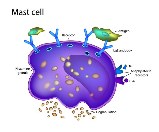
The use of Low-Dose Naltrexone is recent and has been shown to be effective.  Titrating from 1 mg to 4.5 mg daily is typically effective (the maximum dose to remain “low-dose” is 8 mg per day) [[1]].  Low-Dose Naltrexone’s paradoxical effect on the mu-Opioid receptor of increasing the analgesic effect of Opioids has been reported in the literature for decades [[2]].  It also reduces tolerance to Opioids [[3]].  Suppressing the release of proinflammatory substance from Microglia in the Central Nervous System, and inhibition of Mast Cells which uninhibited may release too much Histamine which further stimulates Microglia and other cells in the periphery to release proinflammatory cytokines may be helpful [[4]].  For example, in the gut in the presence of inflamed Endothelium, less Histamine may reduce the inflammation and prevent acid from penetrating into the Stomach wall.  By suppressing Microglia activity, LDN may be very effective in chronic pain in EDS patients with a mix of neuralgic, nociceptive, and neuropathic pain and pain secondary to autoimmune dysfunction.  LDN seems well-tolerated among patients with Fibromyalgia-like symptoms [[5]].  There are many anecdotal reports to support this, but no truly good double-blinded placebo-controlled trial.  Larger trials are needed.  Overall, LDN is a low risk agent to use and may be used in combination with other modalities such as in the treatment of Chronic Fatigue [[6]].  Given that fatigue is often due to Autonomic dysfunction which also promotes inflammation, LDN may have other applications as well.

Some medications, like LDN, have multiple helpful effects by acting on pain neurotransmitters, the endocrine system, and the immune system in beneficial ways. In contrast, Opioids may lower the sensation of pain in the short term but may also sensitize the nervous system and increase pain in the long term. Additionally, while it might be appropriate to take Opioids for pain relief immediately following surgery, for example, if that use progresses to weeks, studies show that continued use one year later is more likely.  Risk of addiction is a significant reason why providers and patients alike are encouraged to explore alternate types of pain management, including exploring any other reasons for amplified perceived pain, including affective disorders.

The role of surrounding muscle for joint connective tissue constraint and protection correlates with the benefits of physical therapy and exercise for treatment of hEDS/HSD [[7],[8]].  Regardless of any other treatments or therapies, hEDS/HSD patients must keep moving, at least in the form of Low-and-Slow exercise.

Lately, our first choice is Low Dose Naltrexone (LDN).  It is not an opioid, nor is it a cannabinoid.  It is, at least in part, a powerful anti-inflammatory which more directly treats the inflammatory responses due to SE amplified by PE.  LDN has proven to be very, very effective and non-addictive at those very low doses.  Reading about it the higher doses states that it has a constellation of symptoms and side effects. But at no more than 8 mg, maybe 10 mg at most, none of those symptoms or side effects are expected.  The only real issue is that it is currently sold only by compounding pharmacies, which may make it inconvenient. If a compounding pharmacy is not known, the local pharmacy will know where a compounding pharmacy would be.  Is recommended if a patient is a chronic NSAID user as a suitable replacement.

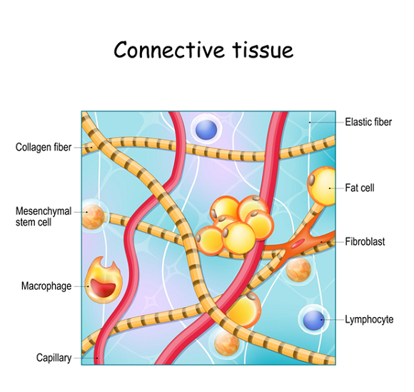
Many hEDS/HSD patients also seek Chiropractic therapy.  In general, it is not recommended.  Traditional Chiropractic therapy will serve to further loosen the already loose joints and may further accelerate the onset of arthritis.  However, there are Chiropractors who are specialized in hEDS/HSD and connective tissue disorders.  These are highly recommended.  Their procedures are more like deep tissue massage to gently realign the joints.

Arthritis is virtually inevitable with hEDS/HSD due to joint laxity.  Rather than gliding smoothly over each other, the bones of the joints tend to bang into each other, causing inflammation.  This inflammation is added to, and amplified by, the inflammation caused by SE; together, they accelerate the onset of arthritis.  Early introduction of natural anti-inflammatories, like LDN or Turmeric or Omega-3 Fatty Acids (if pure), help to reduce inflammation and may help to delay the onset of this arthritis.


[1] Marcus NJ, Robbins L, Araki A, Gracely EJ, Theoharides TC. Effective Doses of Low-Dose Naltrexone for Chronic Pain – An Observational Study. J Pain Res. 2024 Mar 21;17:1273-1284. doi: 10.2147/JPR.S451183. PMID: 38532991; PMCID: PMC10964028.

[2] Gillman MA, Lichtigfeld FJ. A pharmacological overview of opioid mechanisms mediating analgesia and hyperalgesia. Neurol Res. 1985 Sep;7(3):106-19. doi: 10.1080/01616412.1985.11739709. PMID: 2415866.

[3] Wang HY, Friedman E, Olmstead MC, Burns LH. Ultra-low-dose naloxone suppresses opioid tolerance, dependence and associated changes in mu opioid receptor-G protein coupling and Gbetagamma signaling. Neuroscience. 2005;135(1):247-61. doi: 10.1016/j.neuroscience.2005.06.003. PMID: 16084657.

[4] Zhang X, Wang Y, Dong H, Xu Y, Zhang S. Induction of Microglial Activation by Mediators Released from Mast Cells. Cell Physiol Biochem. 2016;38(4):1520-31. doi: 10.1159/000443093. Epub 2016 Apr 7. PMID: 27050634.

[5] Partridge S, Quadt L, Bolton M, Eccles J, Thompson C, Colasanti A, Bremner S, Jones CI, Bruun KD, Van Marwijk H. A systematic literature review on the clinical efficacy of low dose naltrexone and its effect on putative pathophysiological mechanisms among patients diagnosed with fibromyalgia. Heliyon. 2023 Apr 19;9(5):e15638. doi: 10.1016/j.heliyon.2023.e15638. PMID: 37206027; PMCID: PMC10189400.

[6] Bolton MJ, Chapman BP, Van Marwijk H. Low-dose naltrexone as a treatment for chronic fatigue syndrome. BMJ Case Rep. 2020 Jan 6;13(1):e232502. doi: 10.1136/bcr-2019-232502. PMID: 31911410; PMCID: PMC6954765.

[7] Bathen T, Hångmann AB, Hoff M, Andersen LØ, Rand-Hendriksen S. Multidisciplinary treatment of disability in ehlers-danlos syndrome hypermobility type/hypermobility syndrome: A pilot study using a combination of physical and cognitive-behavioral therapy on 12 women. Am J Med Genet A. 2013 Dec;161A(12):3005-11. doi: 10.1002/ajmg.a.36060. Epub 2013 Aug 2. PMID: 23913726.

[8] Castori M. Morlino S.  American Journal of Medical Genetics Part A, 2013; 161, 2989-3004

Read More